
Ulcer depth
Superficial (epithelial) ulcers heal much faster than deep stromal ulcers or those involving Descemet's membrane.
Ulcera corneale · Hornhautgeschwür · Ulcère cornéen · Úlcera corneal
A corneal ulcer is a lesion on the transparent surface of the eye. In the French Bulldog it can appear more easily due to prominent eyes, reduced corneal protection, trauma, dryness, or eyelid abnormalities.
It can cause pain, tearing, redness, a partially closed eye, and opacity. Prompt veterinary assessment is important because some ulcers can deepen rapidly and compromise vision.
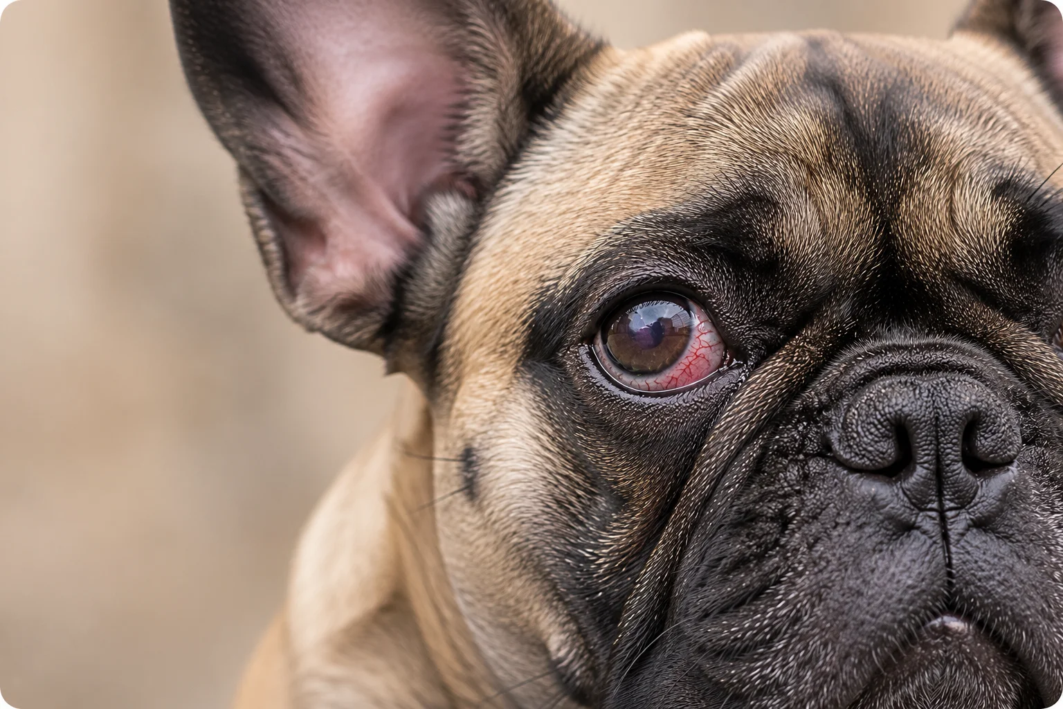
The cornea is the transparent structure that covers the front of the eye and plays a fundamental role in protecting the eye and correctly focusing light.
A corneal ulcer is a loss of tissue from the cornea. It may affect only the most superficial layers or extend deeper into the tissue in more severe cases.
This lesion can appear after minor trauma, contact with foreign bodies, repeated rubbing, alterations of the tear film, or as a consequence of other ocular diseases.
In the French Bulldog the risk is greater because the eyes are more prominent than in other breeds and are therefore less protected from trauma, dust, and dryness.
Some ulcers heal quickly with appropriate treatment, while others can worsen within a few hours or days if not treated promptly.
A corneal ulcer is generally painful. A dog that keeps one eye closed, tears excessively, avoids light, or suddenly develops redness should be seen by a vet promptly.
A lesion of the cornea characterised by loss of the superficial layer or of deeper tissue.
It can be very painful and some forms can worsen rapidly without veterinary treatment.
Partially closed eye, tearing, redness, light sensitivity, rubbing, and possible corneal opacity.
The vet generally confirms the ulcer through an eye examination and fluorescein staining.
Therapy depends on the depth of the ulcer, the cause, and the possible presence of infection.
Prompt veterinary attention reduces the risk of complications and increases the chances of recovery.
Looking at the eye at home does not allow you to determine with certainty how deep a corneal ulcer is. Only a veterinary examination and fluorescein staining can correctly assess its severity.
The French Bulldog has certain anatomical characteristics that make it more exposed to corneal lesions than many other dog breeds.
The eyes are generally larger and more prominent, making them less protected during normal daily activities. Even minor trauma, contact with grass, branches, dust, or simple rubbing can damage the surface of the cornea.
Some individuals may also have eyelid abnormalities, insufficient distribution of the tear film, or other ocular conditions that further increase the risk of developing a corneal ulcer.
Being predisposed does not mean that all French Bulldogs will develop this condition, but knowing the risk factors allows problems to be recognised more quickly and addressed early.
The eyes protrude more than in other breeds and are therefore more exposed to small impacts, foreign bodies, and accidental trauma.
In some French Bulldogs eyelid closure may be less effective, leaving part of the cornea more exposed to the external environment.
Tears protect and nourish the cornea. An insufficient distribution of the tear film can promote irritation and lesions.
Walks in tall grass, play, branches, dust, or simple accidental contacts can cause scratches on the surface of the cornea.
Certain eyelid or eyelash conditions can cause continuous rubbing on the cornea, increasing the risk of ulceration.
The typical conformation of the French Bulldog is one of the main reasons for the greater predisposition to ocular conditions.
Predisposition does not mean that every French Bulldog will develop a corneal ulcer. Regular eye checks and prompt veterinary attention at the first signs help reduce the risk of complications.
Approfondisci → Brachycephalic Obstructive Airway Syndrome (BOAS) in the French Bulldog
The symptoms of a corneal lesion can appear suddenly, sometimes within a few hours, often following trauma, contact with a foreign body, or alterations in the tear film.
Not all ulcers present in the same way. Some are superficial and may improve quickly with appropriate treatment. Others tend to deepen into the inner corneal tissue, increasing the risk of serious complications. The intensity of pain does not always correspond to the severity of the lesion.
Even apparently mild symptoms deserve attention. Early veterinary assessment allows the depth of the lesion to be measured with fluorescein staining and treatment to begin at the most effective time.
In the French Bulldog, prominent eyes make it easier to notice some symptoms, but also increase the likelihood that small corneal lesions worsen rapidly. Early examination can make a difference.
Tap the highlighted points to discover where each symptom appears.
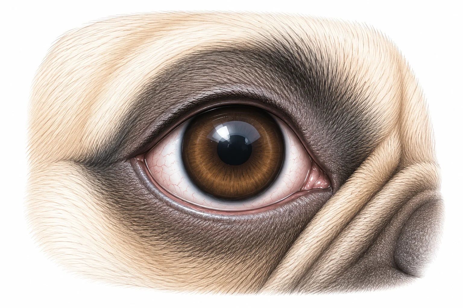
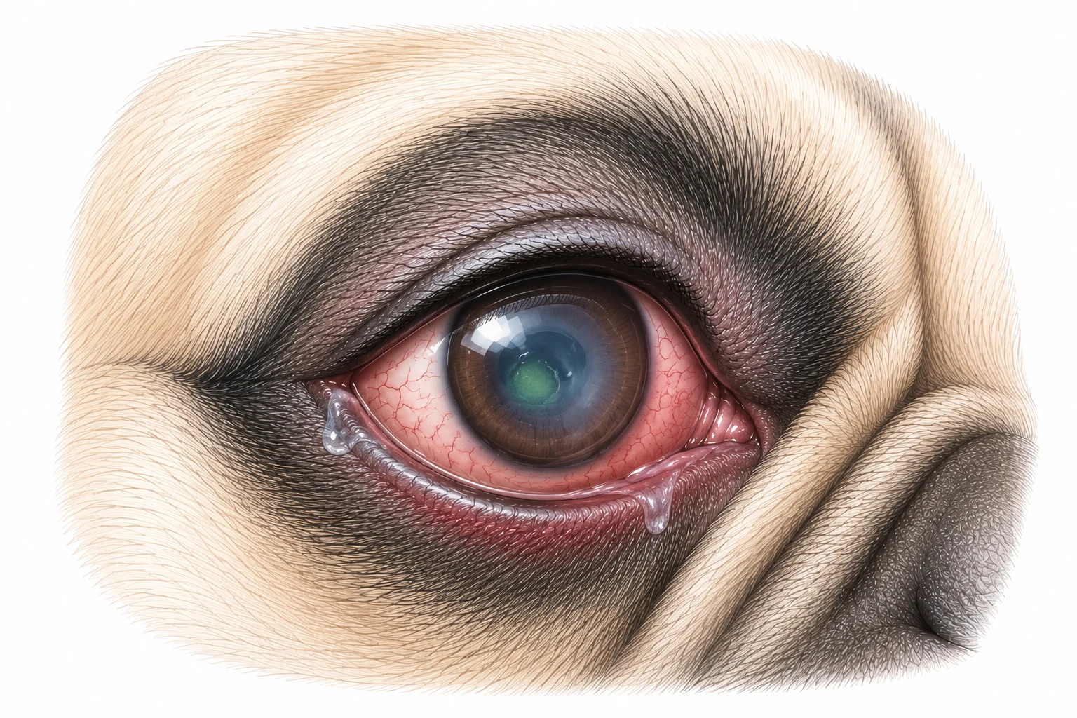
Select a point on the image to see anatomical or clinical details.
The dog tends to keep the eye partially or completely closed due to pain caused by the corneal lesion.
The eye produces more tears than usual in response to irritation and pain.
The cornea may lose its normal transparency, taking on a whitish or bluish appearance.
The conjunctiva and blood vessels of the eye may appear more prominent due to inflammation.
Many dogs seek dimly lit environments or avoid bright light.
The dog may try to rub the eye with its paw or against carpets, furniture, or other surfaces.
Mucous or purulent discharge may appear, especially when a secondary infection is present.
Pain may manifest as restlessness, reluctance to open the eye, vocalisations, or behavioural changes.
Contact your vet immediately if:
Observing your dog's eye can raise suspicion of a problem, but it does not allow you to confirm whether a corneal ulcer is present or to assess its depth. A red conjunctiva, a partially closed eye, or excess tearing can have many different causes.
Only a veterinary visit allows the lesion to be identified, its severity understood, and the most appropriate treatment chosen. There is no need to worry — the examination is quick and generally well tolerated even by anxious dogs.
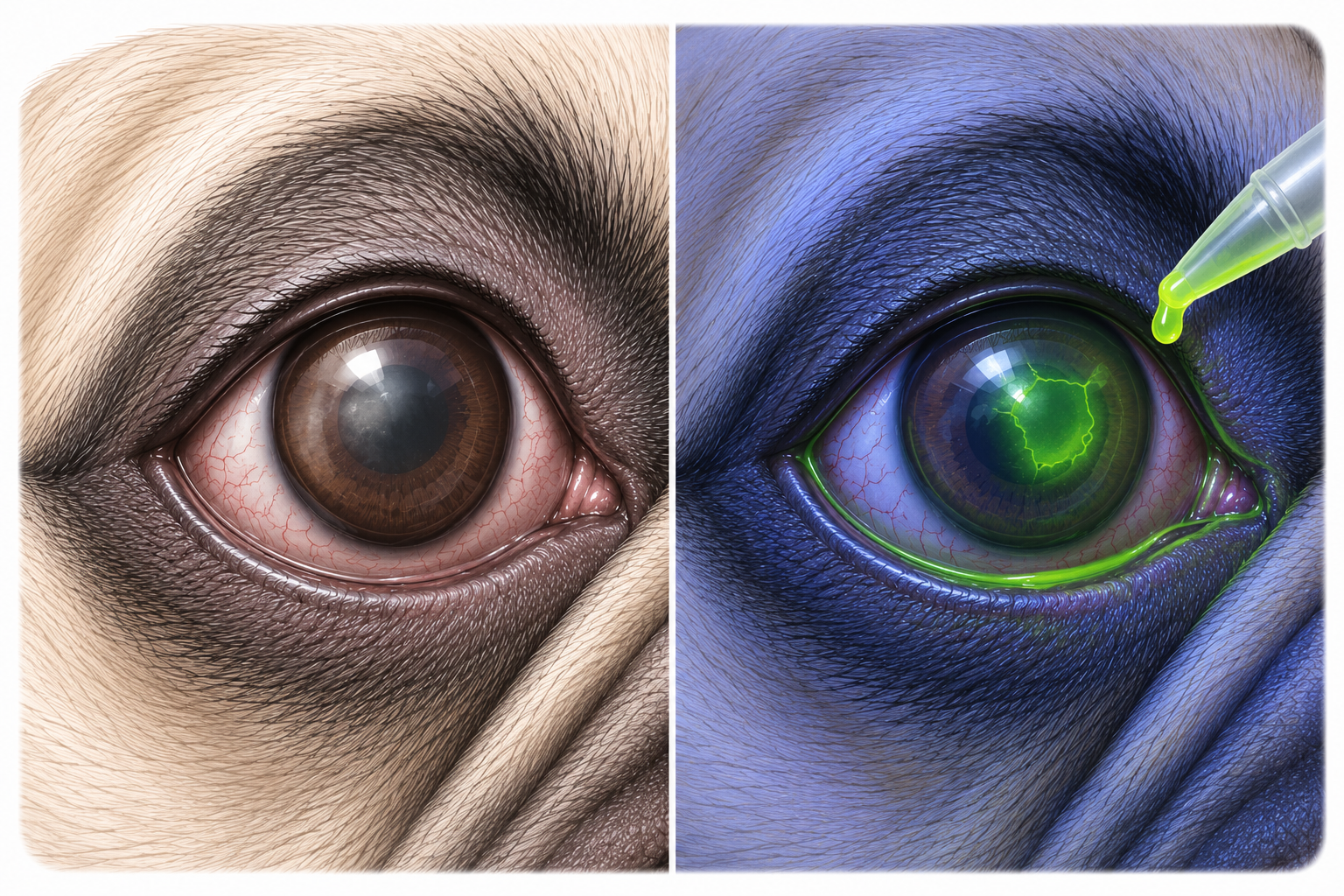
Brief clinical assessment.
Evaluation of cornea, eyelids, conjunctiva, and tear film.
Application of the dye that highlights corneal lesions.
The vet determines whether the ulcer is superficial or deeper.
Treatment is selected based on the results of the examination.
The vet carefully observes the cornea, conjunctiva, eyelids, and other ocular structures to identify any alterations.
A harmless dye is applied to the surface of the eye. Areas where the corneal epithelium is missing become coloured, making the ulcer visible.
The vet assesses potential foreign bodies, eyelid abnormalities, aberrant eyelashes, dry eye, or other conditions that may have caused the lesion.
The depth and extent of the lesion help determine the most appropriate treatment and the frequency of follow-up visits.
The fluorescein test is quick, painless, and is one of the most important tools for confirming the presence of a corneal ulcer.
Treatment depends primarily on the depth of the lesion, the underlying cause, and whether a bacterial infection is present. There is no single protocol: each case is assessed individually by the vet based on the results of the examination.
In superficial cases, medical therapy is generally sufficient and the cornea can heal in a relatively short time with appropriate treatment. In deeper or more complex lesions, a surgical approach may be necessary to protect or repair the corneal structure.
Starting treatment early significantly increases the chances of full recovery and reduces the risk of serious complications, such as corneal perforation or permanent impairment of vision.
The vet assesses the eye with the fluorescein test and determines the severity of the lesion.
The most appropriate therapy is prescribed, which may include topical medications, systemic medications, or both.
The vet repeats the examination to verify the cornea is healing correctly and adjusts treatment if necessary.
In most cases, full recovery is achieved. In more complex cases, surgery may be required.
To prevent or treat bacterial infections when prescribed by the vet.
To reduce pain and improve the dog's comfort during recovery.
Used when necessary to protect the surface of the eye and support healing.
Essential to prevent the dog from scratching or worsening the lesion during treatment.
Foreign bodies, aberrant eyelashes, entropion, dry eye, or other predisposing conditions are treated to prevent recurrence.
Reserved for deep, perforating ulcers or those that do not respond to medical therapy alone.
The vet may repeat the fluorescein test during follow-up visits to check whether the cornea is healing correctly and to decide when to modify or discontinue treatment.
Healing of a corneal ulcer depends on many factors, including the depth of the lesion, the underlying cause, and how promptly treatment was started. Many superficial ulcers can heal completely within a few days when treated early with appropriate therapy.
Deeper ulcers require longer healing times and close monitoring by the vet. In some cases — particularly when a bacterial infection or an anatomical predisposition is present — the healing process can be more complex and may require a surgical approach.
It is important to know that the disappearance of visible symptoms does not necessarily mean the cornea has fully healed. Only a veterinary check-up, with or without a fluorescein test, can verify the actual progression of healing and determine when treatment can safely be discontinued.
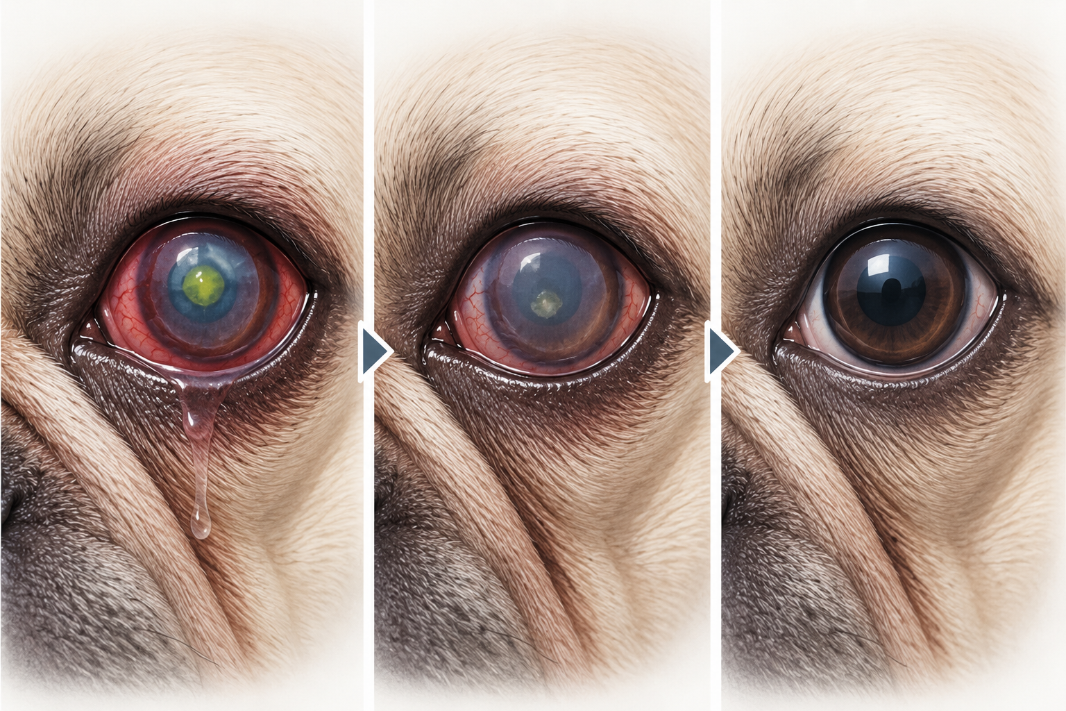
The illustration shows the progressive healing of the cornea: from the presence of the ulcer, to the regrowth of the corneal epithelium, to complete cicatrisation. In more severe cases, a mild corneal scar may remain.
Superficial (epithelial) ulcers heal much faster than deep stromal ulcers or those involving Descemet's membrane.
A superimposed bacterial infection significantly slows healing and increases the risk of serious complications, including melting ulcer and corneal perforation.
Starting treatment early reduces the risk of the lesion deepening and significantly increases the chances of complete healing without residual scarring.
Anatomical factors such as entropion, dry eye (KCS), or abnormal eyelashes can hinder healing if not corrected alongside treatment of the ulcer.
Correct use of the Elizabethan collar is essential: even a single scratching episode can reopen the lesion and compromise healing.
In deep or complicated ulcers, surgery (conjunctival flap, superficial keratectomy) may be necessary, which affects the timing and manner of recovery.
Generally heal within a few days to about a week if treated promptly with appropriate medical therapy.
May require several weeks with frequent veterinary check-ups to monitor the progression of healing.
May require surgical intervention and longer recovery times, with the possibility of residual corneal scarring.
The exact duration varies from case to case: only the vet can provide a realistic estimate based on the specific characteristics of the ulcer.
The disappearance of pain, redness, or tearing does not necessarily mean that the cornea has fully healed. The corneal tissue may appear improved externally, but the lesion may not yet have closed completely. Only a veterinary check-up, often with a new fluorescein test, can confirm complete healing before treatment is safely discontinued.
In most cases diagnosed early and treated appropriately, the prognosis is favourable: many dogs recover full visual function with no permanent effects. In more severe cases, permanent corneal scarring or reduced vision may remain. The speed of veterinary intervention is the factor that most affects the final outcome.
Not all corneal ulcers can be prevented, but many can be avoided by paying attention to the small signals that a dog's eyes send us every day. The French Bulldog is particularly predisposed, due to its brachycephalic conformation which exposes the cornea to a higher risk of trauma and dryness.
Knowing the risk factors and adopting simple daily habits can significantly reduce the likelihood of a corneal ulcer developing, and allow prompt action when the first symptoms appear.
Look for redness, excessive tearing, discharge, or difficulty opening the eye fully. Any abnormality should be assessed by a vet without delay.
During walks in areas with bushes, tall grass, or low branches, the Frenchie's eyes can suffer minor traumas that increase the risk of corneal ulcer.
Prevent the dog from sticking its head out of the window while driving: high-speed wind and debris can cause corneal abrasions and injuries.
In dogs with keratoconjunctivitis sicca (KCS), strictly following the vet-prescribed therapy is essential to keep the cornea adequately lubricated and protected.
Have any eye abnormality checked promptly. In predisposed individuals, periodic ophthalmological check-ups are recommended even in the absence of obvious symptoms.
Conditions such as entropion, abnormal eyelashes (distichiasis, ectopic cilia), or other anatomical defects increase the risk of recurring ulcers and should be surgically corrected.
The brachycephalic conformation of the French Bulldog — with prominent eyes, wide palpebral fissures, and reduced corneal protection — makes these dogs significantly more exposed to eye trauma and corneal dryness than many other breeds. Daily attention to the eyes is particularly important in this case.
Quickly recognising the first symptoms of a corneal ulcer allows, in most cases, treatment to be started early. Prompt action significantly reduces the risk of serious complications and increases the chances of a complete recovery without permanent effects.
A corneal ulcer can worsen very quickly, especially if left untreated. In the French Bulldog, the eye conformation increases corneal vulnerability and can favour rapid progression to deep or complicated lesions.
Do not wait for symptoms to improve on their own: contacting the vet at the first sign of eye discomfort is the safest choice for your dog. Prompt treatment significantly reduces the risk of permanent complications, including loss of vision.
The pain may be intense: an eye kept closed or half-shut indicates a potentially serious lesion requiring immediate assessment.
A cloudy or whitish area on the cornea may indicate significant corneal damage or an ongoing bacterial infection.
Especially if it appears suddenly or is associated with other symptoms, intense tearing can signal a lesion of the eye surface.
Marked redness of the sclera or eyelids is a sign of significant inflammation and requires veterinary assessment without delay.
Repeated scratching or rubbing can rapidly worsen the lesion, turning a superficial ulcer into a deeper one.
Especially if yellowish or greenish, it may indicate a superimposed bacterial infection requiring targeted therapy.
If the dog bumps into objects or seems unable to see well, urgent veterinary assessment is required.
Even if the dog seems slightly better after a few hours, the corneal lesion may continue to worsen without obvious symptoms. Corneal ulcers should never be monitored at home without a veterinary assessment: only a specialist examination — often with a fluorescein test — can correctly evaluate the situation.
Early diagnosis and appropriate treatment significantly increase the chances of complete healing without scarring or vision loss. Every hour of delay can make a difference in the progression of a corneal lesion in the French Bulldog.
Some very superficial lesions may improve, but a corneal ulcer always requires veterinary assessment. Without proper diagnosis and treatment, even a mild lesion can deepen, become infected, and lead to serious complications such as corneal perforation or permanent vision loss.
It depends on the depth of the lesion, the underlying cause, and the response to treatment. Superficial ulcers can heal within a few days with appropriate therapy. Deep or bacterially infected ones may take weeks and, in some cases, require surgery. Only the vet can estimate healing time based on the specific characteristics of the ulcer.
Yes. The brachycephalic conformation — with prominent eyes, wide palpebral fissures, and reduced corneal protection — exposes these dogs to eye trauma, corneal dryness, and associated conditions such as dry eye (KCS) and entropion, all of which increase the risk of corneal ulcer compared with other breeds.
Yes, it is generally very painful. Dogs may hold the eye closed or half-shut (blepharospasm), tear excessively, avoid light (photophobia), and continuously rub their face. The pain is caused by the rich innervation of the cornea, which makes it one of the most sensitive structures in the eye.
Yes. Deep ulcers that have involved the corneal stroma can leave a permanent scar (nebula or leukoma), which in some cases reduces corneal transparency and may partially impair vision. The extent of scarring depends on the depth of the original lesion and the speed of treatment.
No. Most ulcers heal with medical therapy: antibiotic eye drops, lubricants, and pain relief. Surgery is considered for deep, perforating, or medically unresponsive ulcers. The decision is always the vet's after a thorough assessment.
No. Medications must always be prescribed by the vet. In particular, eye drops containing corticosteroids are contraindicated in corneal ulcers: they can slow healing, promote bacterial infections, and significantly worsen the lesion.
Daily eye checks, management of predisposing conditions (entropion, dry eye, abnormal eyelashes), and visiting the vet at the first symptoms are the most effective steps. Avoiding dense vegetation and not letting the dog lean out of the car window are additional useful precautions. In predisposed individuals, periodic eye check-ups are recommended even in the absence of symptoms.
Keep learning about eye health and emergency management in the French Bulldog.
This guide was prepared by consulting veterinary medicine manuals, specialist texts on veterinary ophthalmology, and educational resources from professional organisations.
The content on FrenchieCarePro is for informational and educational purposes only and does not replace a visit, diagnosis, or treatment by a veterinarian. Eye conditions can be painful and deteriorate rapidly: if your dog keeps its eye closed, shows pain, opacity, discharge, marked redness, or has suffered an eye injury, contact a veterinarian promptly. Do not use eye drops, human medications, or unprescribed treatments, especially products containing corticosteroids.
In an emergency, seek immediate veterinary attention or go to an emergency veterinary facility.
Cookie Settings
We use Google Analytics 4 to understand how visitors use this site and improve our content. Analytics cookies are set only if you accept. Declining keeps all features fully functional. Cookie Policy