Patellar Luxation in the French Bulldog
Patellar luxation · Patellaluxation · Luxation de la rotule · Luxación de rótula
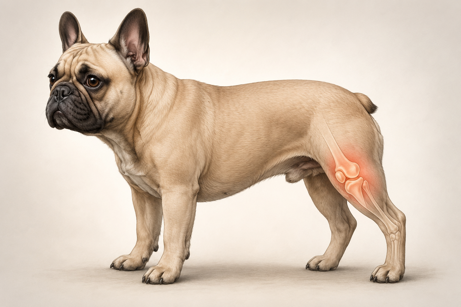
Patellar luxation is an orthopaedic condition in which the kneecap moves out of the femoral groove in which it should glide during knee movement. It may present as intermittent episodes or cause a more persistent alteration in gait.
In the French Bulldog, it is important to recognise early signs such as the characteristic 'skipping' gait, lameness, or intermittent weight-bearing on the hindlimb. The severity of the luxation and its consequences for the joint can vary considerably from one dog to another and must be assessed by a veterinarian.
What is patellar luxation?
The patella is a small bone located at the front of the knee. It is part of the extensor mechanism of the hindlimb together with the quadriceps muscle and the patellar ligament, which connects the patella to the upper part of the tibia.
During normal knee movement, the patella glides within a groove on the distal end of the femur, called the femoral trochlea. This alignment allows the extensor mechanism to function correctly during flexion and extension of the joint.
In patellar luxation, the patella shifts out of the trochlear groove. The displacement may occur towards the inner side of the knee, known as medial luxation, or towards the outer side, known as lateral luxation.
Luxation may occur only under certain conditions or be present for most of the time in more severe cases. Severity cannot be determined simply by looking at how displaced the patella appears in an image: it is assessed by the veterinarian during orthopaedic examination and classified into four grades.
Over time, abnormal patella movement can change the way the structures of the joint work together and, in some dogs, contribute to the development of cartilage changes and degenerative joint disease.
Good to know
Patellar luxation does not necessarily mean the patella is permanently out of place. In milder cases it may displace only occasionally and return to its normal position, while in more severe grades it may remain luxated for most or all of the time.
See what changes in the knee
Compare a normal canine knee with a medial patellar luxation. Tap the highlighted points to learn about the main anatomical structures.
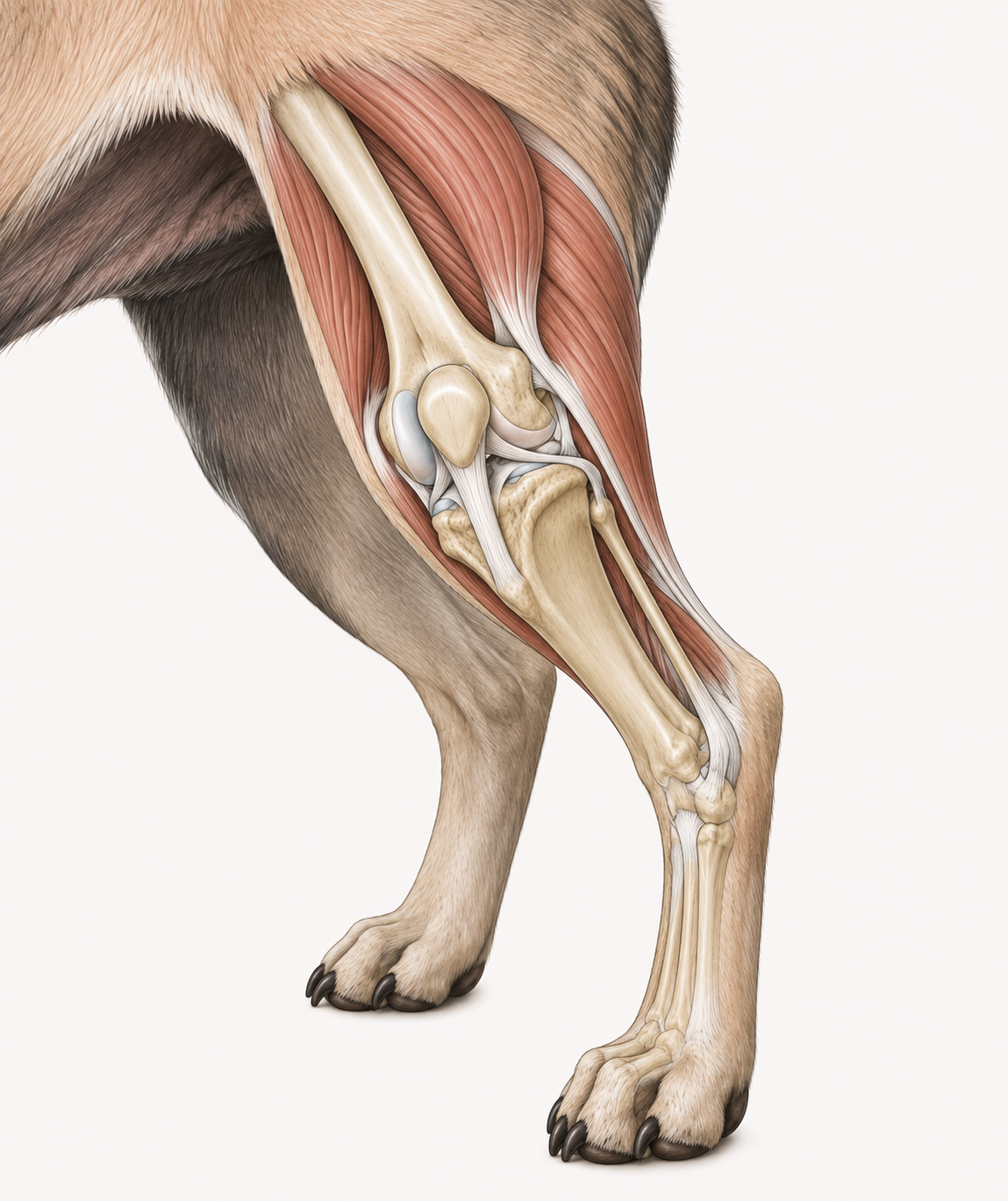
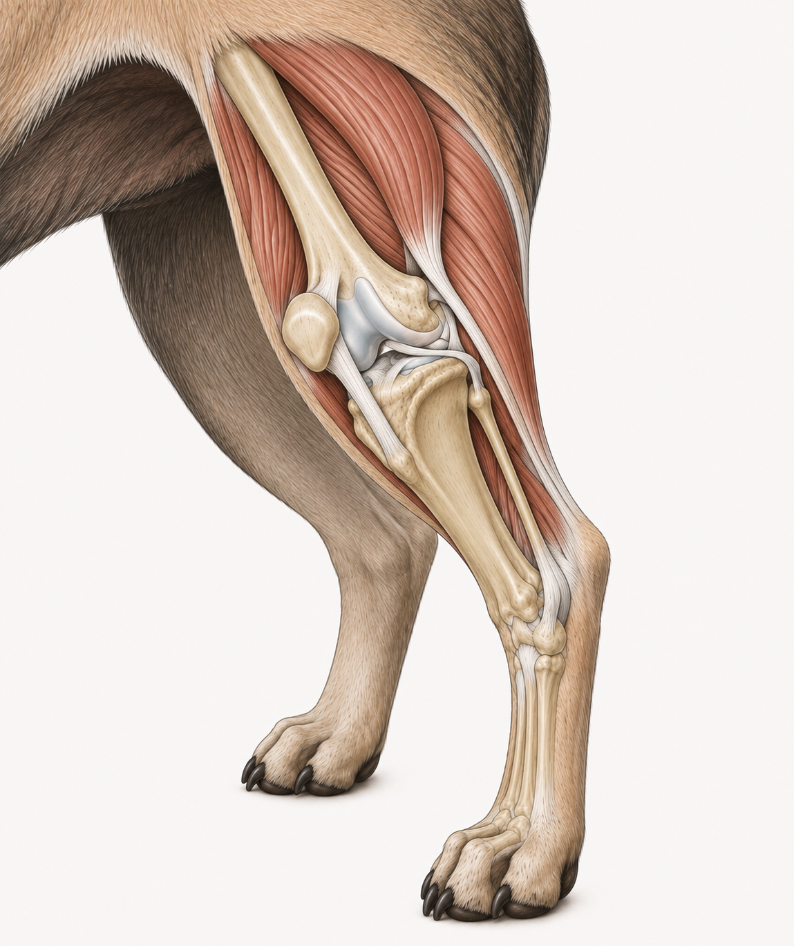
Select a point on the image to discover the anatomical details.
Medial or lateral?
The direction of luxation describes the side towards which the patella shifts relative to the normal femoral groove.
Medial luxation
The patella shifts towards the inner side of the knee.
Lateral luxation
The patella shifts towards the outer side of the knee.
The type of luxation is determined by the veterinarian during orthopaedic examination. This distinction is separate from the grading system, which describes the stability and severity of the luxation.
Remember
The position of the patella in a single image does not allow the grade of luxation to be established. Classification depends on what the veterinarian finds during manipulation of the knee, the ability of the patella to return to the groove, and the presence of any limb deformity.
In Brief
The key points to quickly understand what patellar luxation is, how it may present, and why veterinary assessment is important.
What it is
Patellar luxation occurs when the patella moves out of the femoral trochlear groove. In dogs, the displacement may be medial — towards the inner side of the knee — or lateral, towards the outer side.
Characteristic sign
A frequent sign is the so-called 'skipping' gait: the dog suddenly lifts a hind leg for a few steps, then resumes normal weight-bearing. Symptoms, however, can vary considerably from one dog to another.
Four grades
Patellar luxation is graded by the veterinarian from I to IV, based on patella stability, the ability to displace or reduce it, and the presence of any limb abnormalities.
Diagnosis
Diagnosis is based primarily on orthopaedic examination and palpation of the knee. Radiographs may be used to assess the joint, limb alignment, and any additional changes present.
Treatment
Management depends on the grade of luxation, clinical signs, the dog's age, and the abnormalities present. Some cases can be monitored or managed conservatively, while others may require surgical treatment.
Good to know
The grade of patellar luxation cannot be determined simply by observing the dog's gait or the position of the patella in a photograph. Classification requires an orthopaedic examination during which the veterinarian assesses the position and stability of the patella and the characteristics of the joint.
Why is the French Bulldog predisposed?
Patellar luxation is a developmental orthopaedic condition in the majority of affected dogs. It does not simply result from a single wrong movement: the conformation of the hind limb, the alignment of the knee's extensor mechanism, and a hereditary component are often involved.
In the normal stifle, the patella glides within the femoral trochlear groove as the knee flexes and extends. For this movement to remain stable, the various structures of the limb must be correctly aligned.
In patellar luxation, abnormalities in limb alignment and the extensor mechanism may be present. These variations can alter the direction of forces acting on the patella and predispose it to displacement out of the trochlear groove.
Medial patellar luxation, in which the patella moves towards the inner side of the knee, is a form frequently seen in small-breed dogs and can also affect the French Bulldog.
Predisposition does not mean, however, that every French Bulldog will develop this condition. The presence, severity, and clinical consequences of the luxation can vary considerably from one dog to another.
Limb conformation
The alignment of the femur, tibia, patella, and extensor apparatus influences how the patella tracks within the trochlear groove. Conformation abnormalities can predispose the patella to an abnormal trajectory.
Hereditary component
In most dogs, patellar luxation is considered a developmental condition with a hereditary component. For this reason, orthopaedic evaluation of breeding animals is an important part of responsible selection strategies.
Can affect both stifles
Patellar luxation may be present in one stifle only, or bilaterally. When it is identified in one limb, the veterinarian will therefore generally also evaluate the contralateral knee during the orthopaedic examination.
Predisposition does not mean diagnosis
Being a French Bulldog does not necessarily mean having patellar luxation. Equally, a dog may have a mild luxation without consistently showing obvious signs. Diagnosis and grading must be established through a veterinary orthopaedic assessment.
What about trauma?
Trauma can cause patellar luxation even in a previously normal joint, but this situation is distinct from the developmental form commonly seen in predisposed dogs. In addition, trauma may suddenly make an existing orthopaedic problem apparent.
Symptoms of patellar luxation
The signs of patellar luxation can be highly variable. Some dogs show only occasional episodes of abnormal gait, while others may present more persistent lameness, difficulty in movement, or pain. The severity of the symptoms alone does not determine the grade of the luxation.
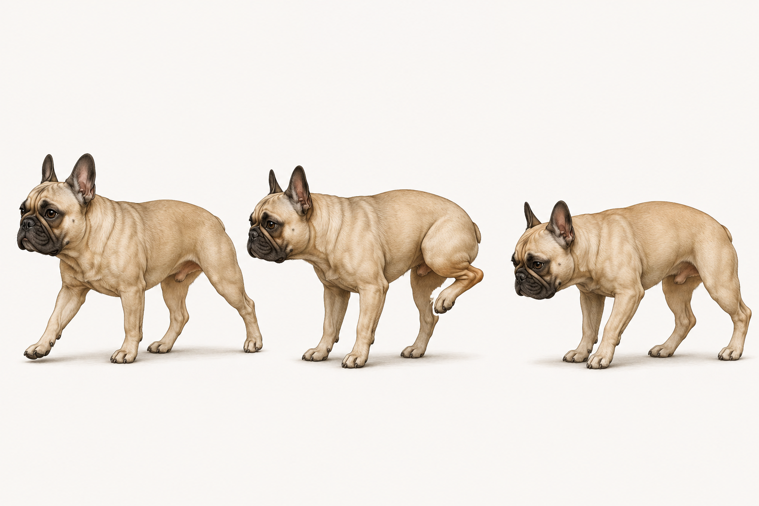
Select a point on the illustration to explore the clinical signs.
Signs to watch for
Patellar luxation does not present in the same way in every dog. Signs may be intermittent or more persistent and can change over time.
- Episodes of skipping during walking or running
- Sudden elevation of a hind limb
- Intermittent or persistent lameness
- Reduced weight-bearing on the affected limb
- Stiffness or changes in gait
- Difficulty or reluctance with certain activities
- Possible pain or discomfort, particularly when associated joint changes are present
Symptoms can vary considerably
A mild luxation may be discovered during a veterinary examination even in a dog with few obvious signs. Conversely, significant lameness does not automatically indicate a high grade of luxation: other orthopaedic conditions can produce similar signs.
For this reason, symptoms and the grade of luxation must be assessed separately by a veterinarian.
When to contact a veterinarian
If your dog shows recurring episodes of skipping, lameness, avoids bearing weight on a paw, or displays persistent changes in gait, it is advisable to schedule a veterinary assessment.
Sudden and marked lameness, intense pain, inability to use the limb, or a significant trauma require prompt veterinary evaluation.
The 4 grades of patellar luxation
Patellar luxation is classified by the veterinarian into four grades, from I to IV. The grade does not simply indicate how displaced the patella appears: it mainly describes its stability, how easily it can leave the trochlear groove, and whether it can be returned to its normal position.
The classification is established through orthopaedic examination. The representations below are a visual simplification intended to help understand the differences between the four grades and cannot replace veterinary assessment.
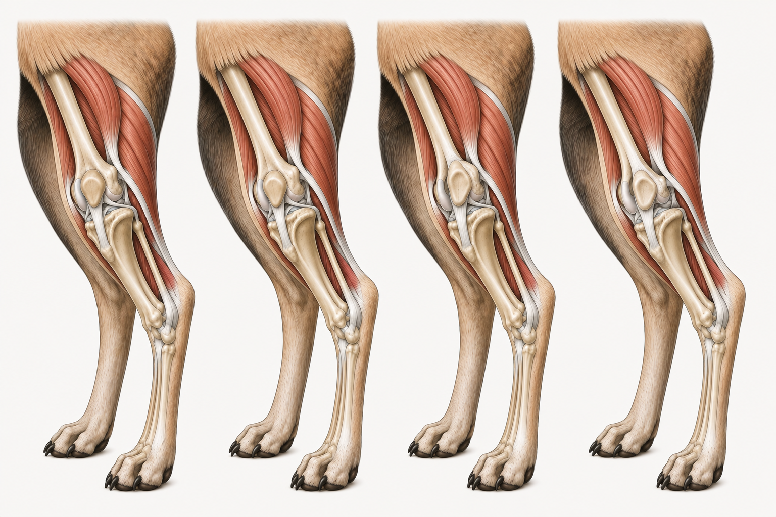
The difference between grades depends not only on how displaced the patella appears, but above all on its stability and the possibility of returning it to the groove during the veterinary examination.
Grade I
Mild instabilityIn grade I the patella is normally located within the trochlear groove. During examination the veterinarian can luxate it manually, but once pressure is released it returns spontaneously to its normal position.
The dog may show no obvious symptoms or may occasionally display brief episodes of abnormal gait.
The grade cannot be determined by observing the dog at home
The frequency of lameness or the severity of symptoms alone do not allow the grade of luxation to be established. Classification requires palpation and manipulation of the joint by the veterinarian, who assesses patellar stability and any associated changes.
If necessary, diagnostic imaging can also help evaluate bone conformation, osteoarthritis, and other stifle abnormalities.
Over time, patellar instability can also affect the other structures of the stifle.
Discover what can happen to the jointHow it can affect the joint
Patellar luxation is not only about the position of the patella. When joint movement remains abnormal over time, the relationships between the surfaces of the stifle may also change, and degenerative changes can develop.
When the patella does not slide normally within the trochlear groove, force distribution across the joint can become abnormal. Repeated contact in non-physiological positions may contribute over time to changes in the articular cartilage and other joint structures.
In some dogs, signs of osteoarthritis may appear — a degenerative joint disease characterised by progressive changes in the cartilage, bone, and tissues involved in joint movement.
The presence and extent of these changes are highly variable. They depend on limb conformation, patellar stability, the duration of the problem, and the individual characteristics of the dog. The grade of luxation and the degree of joint damage are therefore not the same thing.
What can change in the stifle
Compare a relatively normal joint with a representation of chronic changes associated with patellar luxation. The image is educational: the actual severity of changes can vary considerably from one dog to another.
Select a point on the image to find out what can change in the joint.
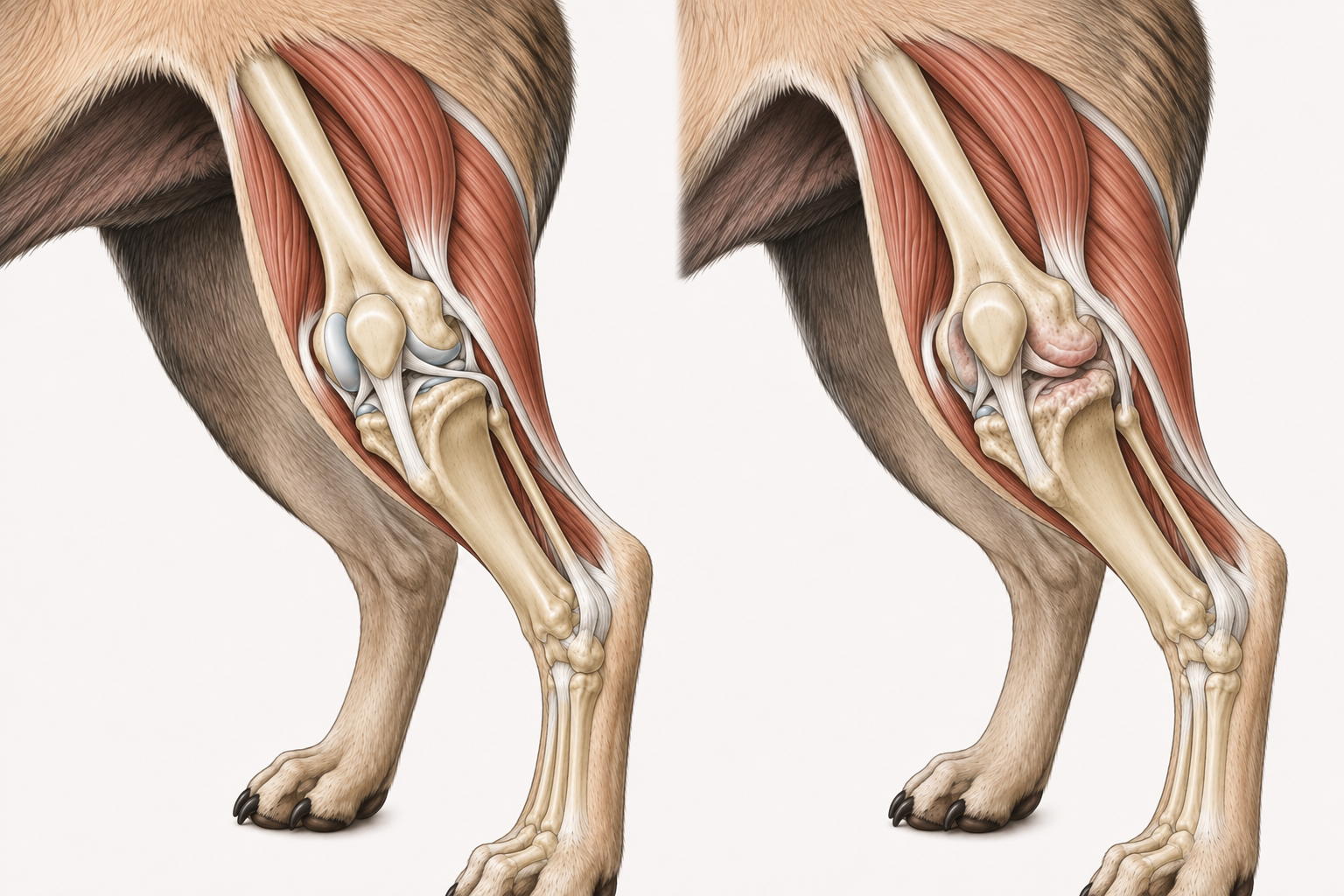
Select a point on the image to find out what can change in the joint.
The illustration shows a didactic comparison. The type and extent of real joint changes can vary considerably.
Luxation and osteoarthritis
Patellar luxation can be associated with pain and osteoarthritis of the stifle. Osteoarthritis is a complex degenerative process that can affect cartilage, subchondral bone, the synovial membrane, and other joint tissues.
Its presence does not depend solely on the grade of luxation. Two dogs with the same grade may present a different joint picture, which is why veterinary assessment must consider the patient as a whole.
Cartilage
Articular surfaces may undergo progressive changes when load and movement are no longer distributed normally.
Bone
With osteoarthritis, bone changes and marginal proliferations may occur, such as osteophytes, which are visible in some cases on radiographic examination.
Pain and function
Joint changes may contribute to pain, stiffness, and reduced limb function, but the intensity and manifestations vary from dog to dog.
Grade of luxation ≠ degree of arthritis
The I–IV classification mainly describes the stability and reducibility of the patella. It does not directly represent the amount of joint damage present. To assess the condition of the stifle, the veterinarian considers orthopaedic examination, symptoms, and — when indicated — diagnostic imaging.
Diagnosis makes it possible to evaluate both the luxation and any associated joint changes.
Find out how it is diagnosedDIAGNOSIS
How is patellar luxation diagnosed?
The diagnosis of patellar luxation is primarily clinical and relies on the orthopaedic examination. The veterinarian observes how the dog walks and uses its hindlimbs, then assesses the knee joint through palpation and specific manipulation manoeuvres.
During the examination, the veterinarian evaluates whether the patella remains within the trochlear groove, whether it can be displaced medially or laterally, whether it returns spontaneously to its normal position, or whether it remains luxated. These findings also contribute to grading the luxation into one of the four severity categories.
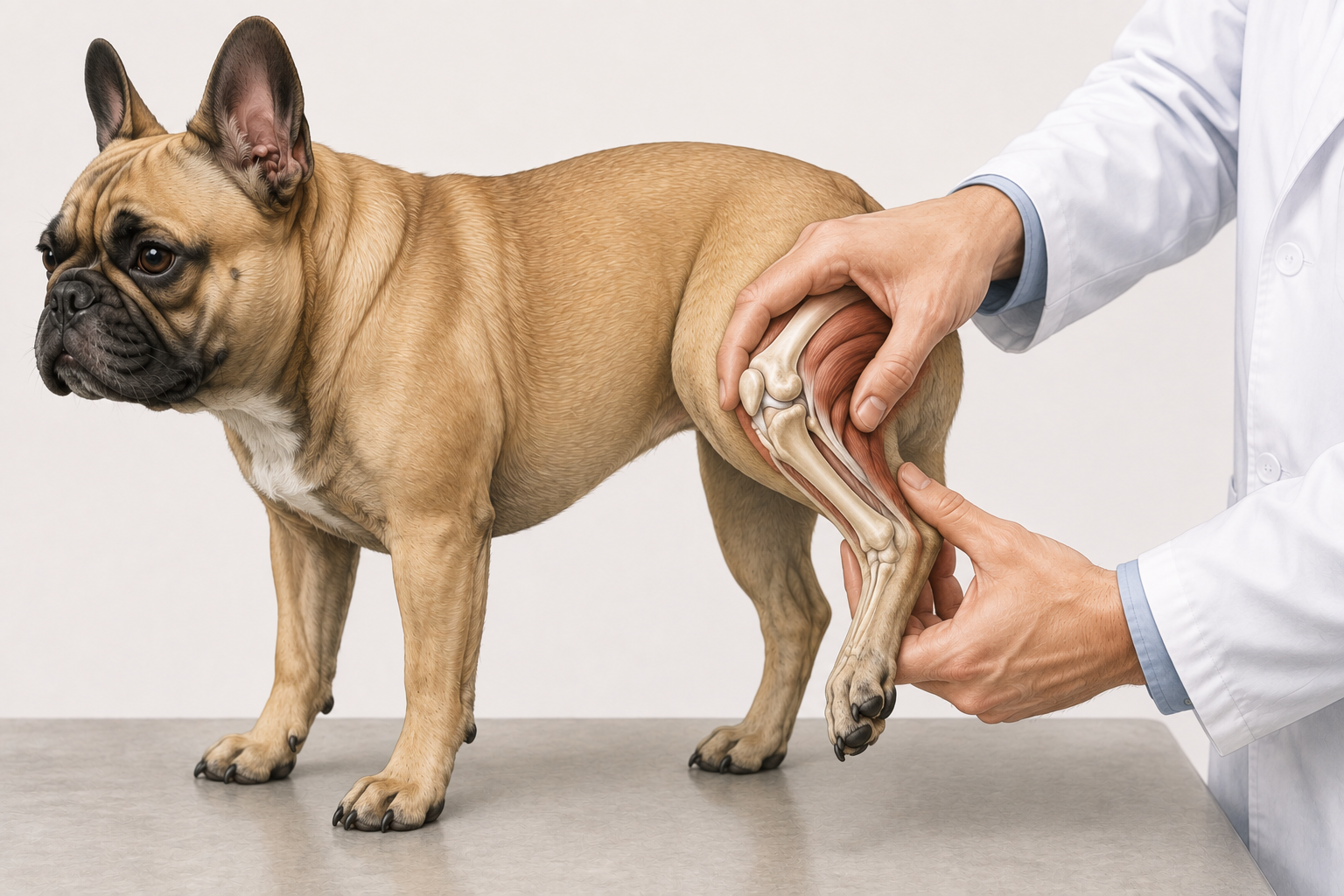
During the orthopaedic examination, the veterinarian manually assesses the position and stability of the patella.
Educational illustration — does not replace a veterinary examination.
How the assessment is performed
Gait observation
The veterinarian observes the dog while it walks and, where useful, during other movements. Intermittent lameness, 'skipping' episodes, reduced weight-bearing on the limb, or persistent gait alterations can provide important information, but they alone are not sufficient to determine the grade of the luxation.
Knee palpation
The patella is palpated during flexion and extension of the knee. The veterinarian assesses its position relative to the trochlear groove and checks whether it tends to shift towards the medial or lateral side of the joint.
Patellar manipulation
Using specific manoeuvres, the veterinarian evaluates whether the patella can be luxated or reduced, whether it returns spontaneously to the normal position, and how easily it tends to luxate again. These characteristics are fundamental for establishing the grade of the luxation.
Assessment of the entire limb
Patellar luxation can be associated with limb alignment abnormalities. For this reason, the veterinarian also evaluates the conformation and function of the entire hindlimb and looks for any other orthopaedic problems that might be contributing to the clinical signs.
Grade is determined by the orthopaedic examination
Observing how often the dog limps is not sufficient to determine whether the luxation is grade I, II, III, or IV. The classification depends mainly on the position of the patella and its behaviour during palpation and manipulation of the joint.
Review the four grades →When are radiographs or other imaging studies needed?
Palpation is central to diagnosing and classifying patellar luxation, but imaging can provide important information about the structure of the joint and the entire limb.
Radiographs
Radiographs can be used to evaluate bone conformation, patellar position, any deformities of the femur or tibia, and the presence of associated joint changes, such as signs of osteoarthritis.
In cases where surgical treatment needs to be planned, radiographic evaluation can also help the veterinarian assess limb alignment.
Computed Tomography (CT)
In more complex cases, particularly when significant femoral or tibial deformities are present, the veterinarian or orthopaedic surgeon may use computed tomography. Three-dimensional images can be especially useful for studying limb anatomy and planning certain surgical procedures.
Not all hindlimb lameness originates from the patella
A dog with patellar luxation may simultaneously have other orthopaedic problems. For this reason, the examination should not focus exclusively on the patella: the veterinarian should evaluate the knee and the entire limb together and, where indicated, investigate other possible causes of the lameness.
How is patellar luxation treated?
Treatment depends on the grade of the luxation, the frequency and severity of clinical signs, the anatomical abnormalities present, and the individual characteristics of the dog. There is therefore no single solution that applies to every patient.
In mild cases or with limited clinical signs, the veterinarian may consider conservative management. When the luxation causes significant clinical problems, is persistent, or significant anatomical abnormalities are present, surgical treatment may be evaluated.
Two possible treatment approaches
Conservative management aims primarily at controlling symptoms and maintaining good limb function. Surgery, when indicated, aims to correct the abnormalities that make the patella unstable and to improve alignment of the extensor mechanism.
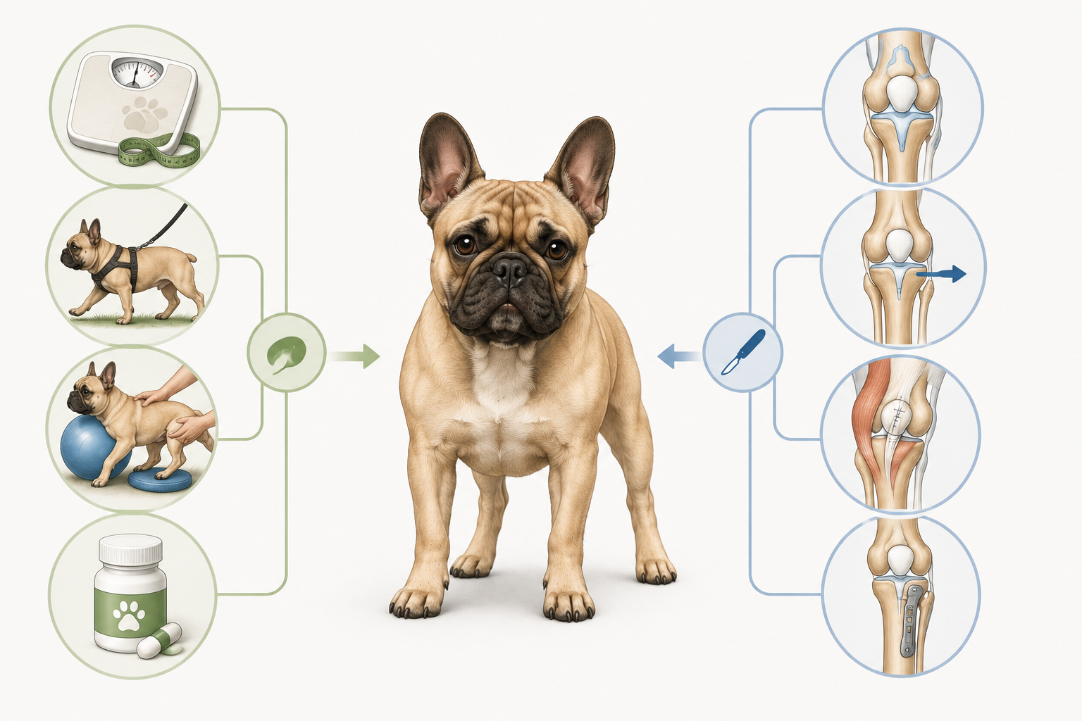
This illustration summarises some treatment principles visually. The actual surgical techniques are chosen and adapted by the orthopaedic surgeon based on the individual dog's anatomy.
Conservative management
In some dogs with mild luxation or limited clinical signs, the veterinarian may decide to monitor the situation and use conservative management.
The goal is to reduce discomfort, maintain good limb function, and limit factors that may increase the load on the joint.
Conservative management does not correct the anatomical abnormalities responsible for the luxation and does not necessarily prevent the condition or joint changes from progressing over time.
Weight management
Maintaining an appropriate body condition reduces the mechanical load on the joints. If the dog is overweight, the veterinarian may recommend a specific nutritional programme.
Controlled activity
Physical activity can be adapted to the dog's condition. The veterinarian may advise regular, controlled exercise while avoiding activities that cause pain or worsen symptoms.
Physiotherapy and rehabilitation
A veterinary rehabilitation programme may be beneficial in some patients to maintain or improve muscle strength, limb control, and joint function. Exercises should be selected according to the individual dog's condition and taught by qualified professionals.
Pain management
When pain or inflammation is present, the veterinarian may prescribe appropriate medications and establish the most suitable therapeutic plan.
What conservative management can and cannot do
Conservative management can help improve comfort and function, but it does not anatomically recentre an unstable patella and does not correct any deformities of the femur, tibia, or extensor mechanism.
For this reason the dog should be re-evaluated over time, especially if lameness becomes more frequent, pain appears, or limb function deteriorates.
Surgical treatment
Surgery is considered when the luxation causes significant clinical signs, is frequent or persistent, or when the anatomical abnormalities make the knee significantly unstable.
The goal is not simply to 'put the patella back in place', but to improve how the patella glides in the trochlear groove and to realign the forces acting on the extensor mechanism.
A single technique is often not sufficient for all cases: the surgeon may combine multiple procedures based on the abnormalities present in each individual dog.
Trochlear groove deepening
When the groove in which the patella glides is too shallow, the surgeon may use trochleoplasty techniques to create a more stable seat for the patella while preserving, as far as possible, the functional articular surface.
Tibial tuberosity transposition
The tibial tuberosity is the point on the tibia where the patellar ligament inserts. In some cases it can be surgically repositioned to improve the alignment of the extensor mechanism and the patellar tracking.
Soft tissue correction
The joint capsule, retinaculae, and other soft tissues can contribute to the forces that keep the patella out of its normal track. In some procedures these tissues are rebalanced to promote better alignment.
Corrective osteotomies
In cases where significant femoral or tibial deformities are present, surgical correction of bone alignment through an osteotomy may be necessary. These procedures are reserved for patients in whom bone deformity contributes significantly to the luxation and require careful orthopaedic planning.
Multiple techniques are often combined
Patellar luxation can result from several anatomical abnormalities occurring simultaneously. For this reason, the surgeon may combine, in the same procedure, techniques on the trochlear groove, the tibial tuberosity, and the soft tissues, or add more complex bone corrections when needed. The choice depends on the anatomy and the needs of the individual patient.
Grade alone does not determine treatment
The grade I–IV classification is important, but the therapeutic decision also considers pain, frequency of luxation, lameness, limb function, age, conformation, osteoarthritis, and any other abnormalities present. Two dogs with the same grade may therefore receive different therapeutic recommendations.
Recovery and prognosis
Recovery after a diagnosis of patellar luxation varies considerably from dog to dog. It depends on the severity of symptoms, the anatomical abnormalities present, the type of treatment chosen, the possible presence of osteoarthritis, and the individual response of the patient.
Many dogs can maintain or recover a good quality of life, but the timeline and functional outcome are not the same for every patient.
When the dog is managed conservatively
In dogs managed without surgery, the aim is to control symptoms, maintain good limb function, and reduce factors that may increase the load on the joint.
The veterinarian may advise activity modifications, weight management, pain control, and, when appropriate, physiotherapy or veterinary rehabilitation.
Improvement in symptoms does not necessarily mean that the patella has become anatomically stable. Luxation may continue to be present even when the dog appears more comfortable.
Improvement in symptoms ≠ anatomical correction
A dog may limp less or move better while still having an unstable patella. For this reason, veterinary check-ups remain important even when symptoms appear to decrease.
After surgical treatment
After surgery, recovery requires a period of limb protection and a gradual return to activity. The precise guidance depends on the techniques used, the dog's individual characteristics, and the surgeon's assessment.
During the postoperative period, clinical check-ups may be scheduled and, when necessary, imaging studies carried out to verify the healing of the treated structures.
Rehabilitation can be introduced progressively when the veterinarian or rehabilitation team considers it appropriate.
How recovery may progress
First weeks
Activity is generally restricted according to the veterinarian's guidance. The initial aim is to protect the knee, manage pain and inflammation, and allow the treated tissues to begin healing.
Gradual resumption of limb use
As recovery progresses, many dogs begin to use the limb more regularly. The pace at which this happens can vary considerably and should not be compared rigidly with other dogs.
Rehabilitation and functional recovery
When indicated, veterinary rehabilitation can help restore muscle strength, mobility, and limb control. The programme is adapted to the stage of healing and the individual patient's condition.
Progressive return to activities
The return to longer walks, play, and other activities is decided gradually by the veterinarian based on knee stability, healing progress, and limb function.
How long does recovery take?
There is no single timeframe that applies to every dog.
In straightforward cases, the veterinarian may observe functional improvement within a matter of weeks, whereas after more complex procedures or when significant functional deficits are present, the process may take several months.
These are guidelines only. The most important parameter remains the clinical progress of the individual dog and the assessment of the veterinary team.
What determines the prognosis?
Prognosis does not depend on a single factor. The final outcome is influenced by the combination of clinical severity, limb anatomy, any articular changes present, and the response to treatment.
Severity of symptoms
The frequency of lameness, the presence of pain, and the degree of functional impairment influence the therapeutic course and recovery.
Anatomical abnormalities
Deformities of the femur or tibia, alignment of the extensor mechanism, and the conformation of the trochlear groove can make the clinical picture more complex.
Joint damage
The presence of osteoarthritis or other degenerative changes can affect comfort and function even after treatment of the luxation.
Treatment performed
Recovery also depends on the type of management adopted and, in surgical cases, on the procedures required to correct the abnormalities present.
Individual response
Age, physical condition, body weight, healing capacity, and adherence to post-treatment guidance can all contribute to the final outcome.
Many dogs can maintain a good quality of life
With appropriate management, many dogs with patellar luxation can live with good function and a good quality of life.
Some recover very satisfactory function, while others may retain a degree of stiffness, intermittent lameness, or chronic joint changes.
The outcome depends on the individual case and should be assessed over time.
Can the luxation recur?
After surgical correction, patellar stability may improve significantly, but no treatment can guarantee that knee problems will never occur again.
Joint changes may persist or develop over time, and in some dogs instability, lameness, or other orthopaedic problems may reappear.
For this reason, follow-up appointments and observation of gait remain important even after a good recovery.
When to seek veterinary reassessment
Contact your veterinarian if any of the following occur during recovery:
- Sudden worsening of lameness
- Obvious pain
- Persistent refusal to use the limb
- Significant swelling
- Deterioration after a period of improvement
- New difficulties with movement
Case study: Zeus
A real journey from diagnosis to recovery
Zeus is a French Bulldog diagnosed with grade III medial patellar luxation in both knees. Because the left hindlimb showed more evident clinical signs, the left knee underwent surgical treatment.
His story provides a concrete example of how diagnosis, surgery, rehabilitation and follow-up examinations can all be part of the same treatment pathway.
This section describes a single case.
An individual case, not a model for all
Zeus's journey does not represent what necessarily happens to every dog with patellar luxation. Surgical indications, techniques used, complications, recovery times and the final outcome can differ in each patient.
This case is presented solely for educational purposes.
6 November 2025
Veterinary documentationBilateral grade III medial luxation
Veterinary documentation records grade III medial patellar luxation bilaterally.
At the time of assessment, Zeus was weight-bearing on the left hindlimb significantly less often than on the right.
Surgical treatment was therefore performed on the left knee.
LEFT KNEE
Limb treated surgically
BOTH KNEES
Grade III medial luxation
6 November 2025
Veterinary documentationThree procedures in a single operation
To correct the luxation of the left knee, multiple surgical techniques were used in the same procedure:
- Sulcoplasty
- Tibial tuberosity transposition
- Lateral imbrication
This is a concrete example of how the correction of patellar luxation may require a combination of procedures tailored to the anatomical abnormalities present in the individual knee.
6 November 2025
Veterinary documentationCartilage changes already present
During the procedure, mild synovitis and mild hypertrophy of the synovial villi were documented.
The operative report also describes grade III chondromalacia according to the Modified Outerbridge Score affecting the patella, predominantly medially, and the involved medial femoral surface.
The cranial and caudal cruciate ligaments and the menisci were intact.
What does chondromalacia mean?
Chondromalacia refers to an alteration of the articular cartilage. In Zeus's case, the finding documents that, in addition to patellar instability, changes to the cartilage surface were already present.
After the procedure
Veterinary documentationLimb protection and controlled movement
The postoperative plan included short controlled walks at a slow pace, 5 to 10 minutes, 3–5 times a day, on a short lead.
During this initial phase, free running, jumping, vigorous play and stairs were to be avoided until the next reassessment.
Wound checks, an orthopaedic review and a radiographic check were also planned according to the schedule indicated by the veterinary facility.
These were the instructions given specifically to Zeus. They are not a universal protocol applicable to all dogs.
Postoperative period
Owner-reportedRehabilitation during recovery
During the postoperative period, Zeus also underwent professional physiotherapy.
The programme included:
- Massage
- Laser therapy
- Rehabilitation exercises
- Underwater treadmill
The clinic's documentation expressly recommended professional physiotherapy in the postoperative period. The specific rehabilitation modalities described here are those reported by the owners.
During recovery
Owner-reportedReturn to normal gait
During recovery, Zeus had returned to walking normally without limping and without displaying the characteristic 'skipping' gait that can accompany patellar luxation.
12 January 2026
Veterinary documentationAn implant-related complication
An implant reaction subsequently developed in relation to the hardware used during the tibial tuberosity transposition.
The radiographic check reported that the osteotomy had healed well, while soft-tissue swelling was present around the implant exit site.
Implant removal was therefore decided upon.
The report describes an implant reaction, not an infection, rejection or allergy. This does not constitute a failure of the original procedure.
12 January 2026
Veterinary documentationImplant removal
Zeus underwent implant removal. According to the report, anaesthesia and the procedure proceeded without complications and Zeus was discharged in good general condition with postoperative medication.
A sample was also sent for bacteriological examination.
The following day
Owner-reportedCheck-up with the regular veterinarian
The day after implant removal, Zeus was seen by his regular veterinarian to check the wound and assess his general condition following anaesthesia.
According to the owners, the check-up revealed no problems.
Approximately two weeks later
Owner-reportedSuture removal
After approximately two weeks, the sutures were removed. No further interventions were required in relation to this phase of recovery.
According to the owners' observations, Zeus was walking normally and showed no lameness or skipping episodes.
Zeus's radiographs
These are real radiographs of Zeus's left knee taken during his clinical journey.
Implant still in place
Radiograph of the left knee with the osteosynthesis material still present after tibial tuberosity transposition.
After removal
Radiograph of the left knee after implant removal.
Real radiographs from Zeus's clinical case, published with the owner's consent.
What the report documents
At the time of implant removal, the radiographic report described the osteotomy as well healed. The decision to remove the hardware was linked to the implant reaction and the soft-tissue swelling in the area involved.
What Zeus's case shows us
The same grade does not mean the same symptoms
Zeus had a grade III luxation in both knees, but offloaded the left limb more frequently than the right.
Surgery may combine multiple techniques
In the left knee, sulcoplasty, tibial tuberosity transposition and lateral imbrication were performed together.
Recovery does not end with the operation
Follow-up checks, controlled activity and rehabilitation were all part of Zeus's postoperative journey.
Complications can occur
In Zeus's case, an implant reaction made subsequent removal necessary, despite the osteotomy being well healed.
An individual outcome
In Zeus's case, the journey ended with a return to normal gait without lameness or skipping observed by the owners.
This outcome does not allow prediction of the course in another dog. Every patellar luxation must be assessed individually by a veterinarian on the basis of symptoms, orthopaedic examination and the anatomical abnormalities present.
Can patellar luxation be prevented?
Not always.
Patellar luxation is often linked to limb conformation and the development of the extensor mechanism. When an anatomical predisposition is present, the owner cannot eliminate it simply by modifying the dog's activity.
Appropriate management can, however, help reduce the load on the joints, maintain good muscle function and detect any changes in gait at an early stage.
Maintaining a healthy body weight
Being overweight increases the mechanical load on the joints. Maintaining an appropriate body condition can help reduce stress on the knee and protect joint function over time.
If the dog is overweight, a veterinarian can advise on a suitable nutritional plan.
Regular, appropriate physical activity
Regular physical activity suited to the dog's condition contributes to maintaining muscle mass and limb function.
In dogs with a confirmed diagnosis or orthopaedic symptoms, the type and intensity of activity must be adapted according to the veterinarian's guidance.
Reducing slipping and uncontrolled movement
Very slippery surfaces can make it harder for the dog to control the limb and may increase the risk of sudden movements or loss of balance.
Rugs, non-slip surfaces or other home adaptations can be helpful, especially in dogs that already show instability, lameness or difficulty moving.
Watching for changes in gait
Recurrent 'skipping', sudden lifting of a hindlimb, lameness or persistent changes in weight-bearing deserve attention.
Recognising these signs early allows the dog to be assessed before any joint problems become more significant.
Veterinary check-ups
In dogs with a confirmed diagnosis, veterinary check-ups allow symptoms, patellar stability, limb function and any associated joint changes to be monitored.
Even in dogs without a diagnosis, an orthopaedic assessment is indicated when recurrent or persistent signs appear.
Stronger muscles do not correct anatomy
Good muscle condition can contribute to limb function and control, but it does not correct femoral or tibial deformities, a shallow trochlear groove or abnormal alignment of the extensor mechanism.
Physiotherapy and controlled activity can be important tools in management, but they do not 'put back in place' a patella predisposed to luxation.
Ramps, steps and the home environment
Ramps, low steps, non-slip surfaces and other adaptations can be helpful for some dogs, particularly when pain, instability or difficulty moving is present.
These tools should be considered as supports to reduce sudden movements or make daily life easier — not as methods capable of preventing or correcting patellar luxation.
What about responsible breeding?
Patellar luxation is considered, in most dogs, a developmental condition with a hereditary and conformational component.
For this reason, orthopaedic evaluation of breeding animals is important in responsible selection strategies.
A dog affected by patellar luxation should be carefully evaluated by a veterinarian before being used for breeding.
What the owner can do in practice
- maintain the dog in a good body condition
- encourage regular and appropriate physical activity
- watch for any changes in gait
- seek veterinary assessment when recurrent or persistent symptoms appear
These measures can contribute to joint health and the dog's management, but they do not eliminate an anatomical predisposition that is already present.
WHEN TO SEEK VETERINARY CARE
When should you consult a veterinarian?
An occasional episode of abnormal limb movement alone is not enough to confirm the presence or severity of patellar luxation. However, when lameness, recurrent 'skipping', pain or persistent changes in gait appear, it is advisable to have the dog assessed by a veterinarian.
An orthopaedic examination allows the stability of the patella to be checked and helps distinguish patellar luxation from other possible causes of hindlimb lameness.
Schedule an appointment
It is advisable to arrange a veterinary assessment when you notice one or more of these changes:
- recurrent 'skipping' episodes during walking or running
- intermittent lifting of a hindlimb
- lameness that comes and goes
- persistent changes in the way the dog walks
- reduced willingness to engage in activities that were previously performed normally
- suspected patellar luxation even in the absence of obvious pain
The frequency of symptoms alone does not determine the grade of the luxation.
Contact your veterinarian promptly
A more timely assessment is indicated when:
- lameness noticeably worsens
- the dog shows obvious pain
- the limb is used much less than usual
- difficulty moving persists rather than resolving
- obvious swelling appears around the knee
- symptoms worsen after a previous period of improvement
- an operated dog shows a significant change in limb use
When urgent assessment is needed
Some signs are not typical of simple intermittent patellar luxation and require urgent veterinary assessment, particularly if they appear suddenly.
- sudden inability to use a limb
- intense or difficult-to-control pain
- significant trauma followed by severe lameness
- obvious deformity of the limb
- significant wound or bleeding in the affected area
- rapid deterioration of general condition alongside the orthopaedic problem
These signs may have causes other than patellar luxation. Veterinary assessment is necessary to determine what is happening.
If the dog has had surgery
During postoperative recovery, it is important to follow the instructions provided by the surgeon and attend scheduled check-ups.
Contact the veterinary team if you notice significant worsening of lameness, increasing pain, marked swelling, wound problems or an unexpected change in the recovery progress.
What to do while waiting for the appointment
If the dog is in pain or lame, keep it calm and limit activities that provoke or worsen symptoms until the veterinary assessment.
Avoid repeatedly manipulating the knee in an attempt to check or 'put back' the patella.
Do not administer human medicines or other medications without veterinary guidance.
If the dog cannot use the limb, is in severe pain or has suffered significant trauma, seek timely veterinary assistance.
FREQUENTLY ASKED QUESTIONS
Frequently asked questions about patellar luxation
No. Some dogs may have patellar luxation with mild or intermittent symptoms, while others develop more obvious lameness, discomfort or pain.
The presence and intensity of pain depend on the individual case and may be influenced by the frequency of luxation and associated joint changes.
No. The decision does not depend solely on the presence of luxation or its grade.
The veterinarian considers symptoms, frequency of episodes, pain, limb function, luxation grade, anatomical conformation, any joint changes and the individual dog's characteristics.
Some patients can be managed conservatively, while in others surgical treatment is recommended.
No. Grade III indicates a significant luxation, but the classification alone does not replace a clinical assessment.
The treatment decision also takes into account symptoms, limb function, anatomical abnormalities, age, general condition and other orthopaedic findings.
Yes. Patellar luxation can be bilateral, affecting both knees.
Clinical severity can, however, differ between the two limbs, even when the assigned grade is the same.
Zeus's case study on this page is an example: veterinary documentation recorded bilateral grade III medial luxation, but the left limb was offloaded more frequently.
Read Zeus's case →Physiotherapy can help improve muscle strength, limb control and function, but it does not correct bone deformities or other anatomical abnormalities responsible for the luxation.
When indicated, rehabilitation must be tailored to the individual dog's condition by qualified professionals.
In many cases yes, but the type and intensity of activity must be adapted to the dog's condition.
There is no universal rule prohibiting running, play, stairs or other activities for all dogs with patellar luxation.
When symptoms are present, after an operation or in more complex cases, it is the veterinarian who determines appropriate restrictions.
No. A ramp cannot correct or eliminate an anatomical predisposition to luxation.
It may, however, be useful in some dogs to reduce jumping or make access to certain surfaces easier, especially when pain or difficulty moving is already present.
Being overweight increases the load on the joints and can make managing an orthopaedic problem more difficult.
Maintaining an appropriate body condition is therefore an important part of joint health, but weight loss does not correct the anatomy responsible for the luxation.
In some dogs the picture may remain relatively stable, while in others the frequency of symptoms, instability or joint changes may increase.
The progression is not the same in all patients. For this reason, once luxation has been diagnosed, clinical monitoring over time can be important.
Surgery aims to improve patellar stability and extensor mechanism alignment, but no procedure can guarantee the complete absence of future problems.
The outcome depends on the severity of the case, anatomical changes, procedures performed, healing and other individual factors.
The grade is determined primarily during the orthopaedic examination through palpation and manipulation of the patella.
The veterinarian assesses the position of the patella, whether it can be luxated or reduced manually, whether it returns spontaneously to the groove and how stably it remains there.
Radiographs can provide important information about bone structures and associated changes, but do not replace the orthopaedic examination in clinical grading.
No. The characteristic intermittent lifting of a hindlimb may be consistent with patellar luxation, but alone it does not allow a diagnosis to be made.
Other orthopaedic problems can cause gait abnormalities. Diagnosis requires veterinary assessment.
In most dogs, patellar luxation is considered a developmental condition with a hereditary and conformational component.
Transmission should not, however, be described as dependent on a single gene or as completely predictable.
Orthopaedic evaluation of animals intended for breeding is therefore important within the framework of responsible selection.
It is useful to observe how the dog walks and note any episodes of lameness or 'skipping', but it is not advisable to repeatedly manipulate the knee in an attempt to provoke or correct a luxation.
Palpation and grading of the patella must be performed during a veterinary orthopaedic examination.
Related symptoms
Rehabilitation
Other diseases in the same category
SOURCES & REVIEW
Veterinary sources
The content of this page was developed using authoritative veterinary sources and relevant scientific literature, with the aim of providing comprehensible information based on available veterinary knowledge.
The information is for educational purposes and does not replace a veterinary examination, diagnosis or treatment.
Merck Veterinary Manual
Patellar Luxation in Dogs and Cats
Veterinary overview of aetiology, clinical signs, diagnosis and treatment of patellar luxation in dogs and cats.
(opens in a new tab)American College of Veterinary Surgeons (ACVS)
Patellar Luxations
Specialist information on patellar luxation, clinical assessment and main surgical treatment options.
(opens in a new tab)VCA Animal Hospitals
Luxating Patella in Dogs
Veterinary resource covering symptoms, diagnosis, grading and management of patellar luxation in dogs.
(opens in a new tab)Orthopedic Foundation for Animals (OFA)
Patellar Luxation
Information on orthopaedic assessment of the patella and the role of screening in responsible breeding selection.
(opens in a new tab)Scientific literature
The content of this page also draws on relevant peer-reviewed veterinary literature on patellar luxation.
Case study documentation
Zeus's case study presented on this page is based on the original veterinary documentation relating to his clinical journey and on subsequent observations reported by the owners.
- veterinary report dated 06.11.2025
- documentation relating to the surgical procedure of 06.11.2025
- postoperative treatment plan
- veterinary report dated 12.01.2026 relating to the implant reaction and removal
- original radiographs from the case
- subsequent course as reported by the owners
Educational information, not a diagnosis
FrenchieCarePro provides informational and educational content dedicated to the health and wellbeing of the French Bulldog.
The information on this page cannot replace a clinical examination, diagnosis or therapeutic guidance from a veterinarian.
If your dog shows pain, lameness, difficulty moving or other symptoms, consult a veterinarian.
If symptoms are sudden or severe, seek timely veterinary assistance.
Last review: August 2026