IVDD (Intervertebral Disc Disease) in the French Bulldog
Intervertebral Disc Disease • Bandscheibenerkrankung • Hernie discale • Hernia discal
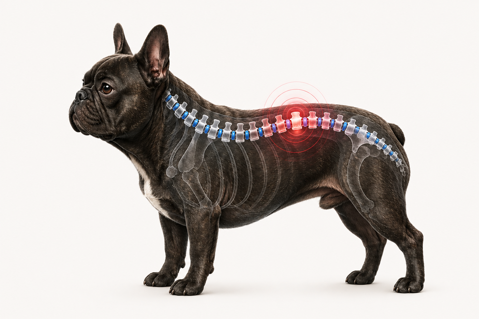
IVDD (Intervertebral Disc Disease) is one of the most common neurological conditions in the French Bulldog. When an intervertebral disc is damaged, it can compress the spinal cord, causing pain, difficulty walking and, in severe cases, paralysis. Recognising the early signs and acting quickly can significantly improve the prognosis.
What is IVDD?
The spine consists of a series of vertebrae. Between most of them are intervertebral discs — elastic structures that absorb some of the forces on the spine and allow it to move.
Each disc is made up of a tougher outer layer called the fibrous annulus, and a softer inner part called the nucleus pulposus. The disc sits close to the vertebral canal, through which the spinal cord passes.
The term IVDD, from the English Intervertebral Disc Disease, refers to a degenerative condition of the intervertebral discs. The disc may lose elasticity, become dehydrated, calcify, or weaken, making it more vulnerable to protrusion or rupture.
When part of the disc protrudes or its inner material leaks into the vertebral canal, it can compress the spinal cord or nerve roots. This condition is more precisely defined as intervertebral disc herniation and can cause pain and neurological changes of varying severity.
The consequences depend on the position of the affected disc, the amount of extruded material, the force and duration of compression, and the damage sustained by the spinal cord. Some dogs mainly experience pain, while others may develop weakness, difficulty walking, or paralysis.
See what changes in the disc
Tap the buttons and highlighted points to compare a healthy intervertebral disc with a disc that compresses the spinal cord.
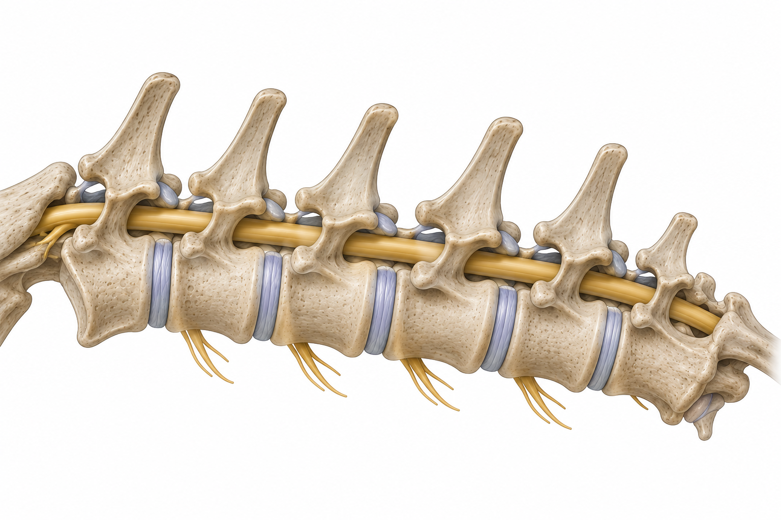
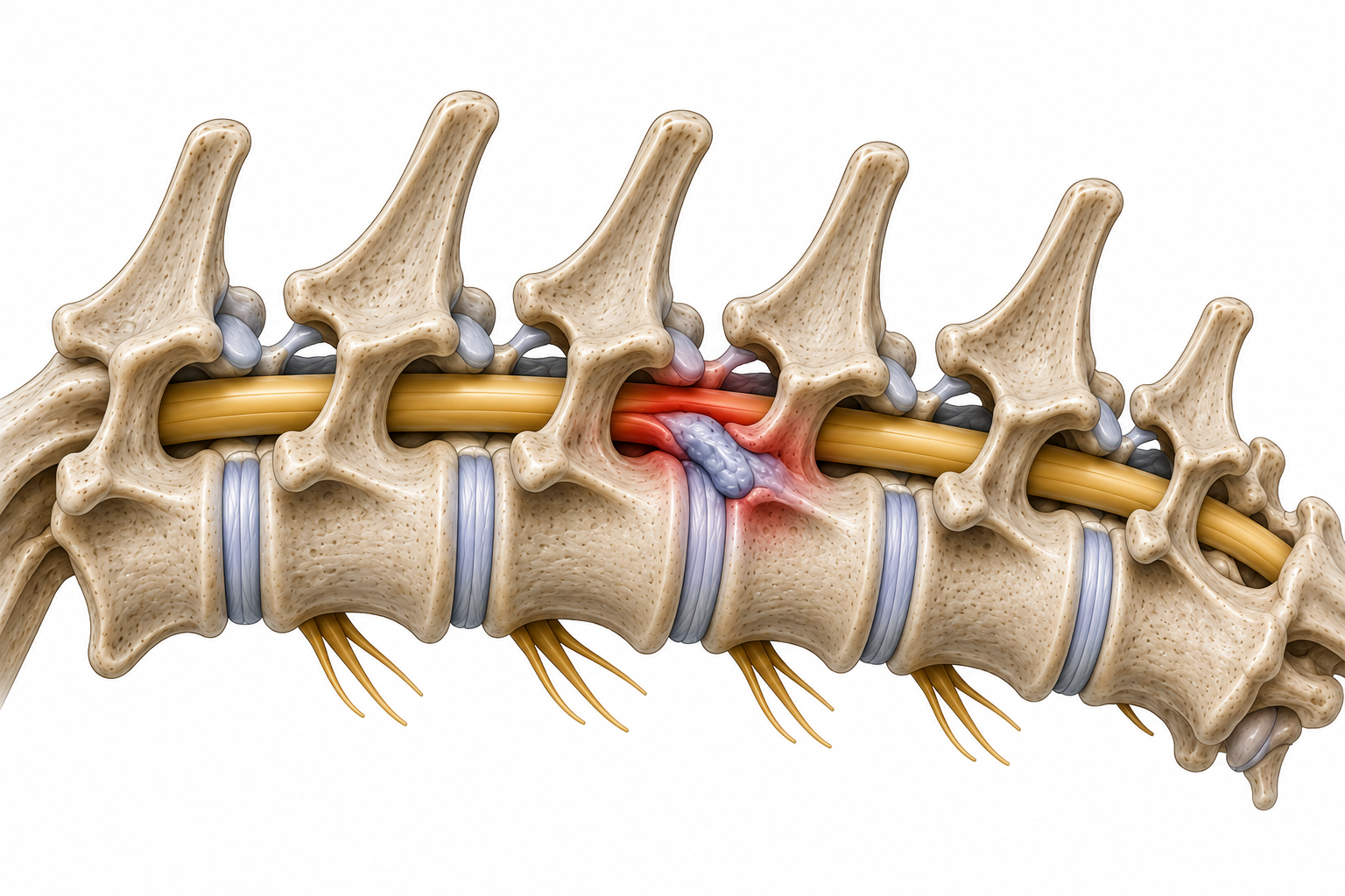
Select a point on the image to see anatomical details.
Good to know
IVDD and disc herniation are not perfectly identical terms. IVDD refers to the degenerative process that weakens the disc; herniation occurs when the disc protrudes or its material leaks out and may compress the spinal cord. In everyday and educational language, both terms are often used together.
At a glance
What it is
A degenerative disease of the intervertebral discs. When a disc deforms or its material leaks into the vertebral canal, it can compress the spinal cord or nerve roots.
Why it matters
Compression of the spinal cord can cause pain and neurological deficits of varying severity. In more serious cases, the dog may lose the ability to walk or develop paralysis.
Common signs
Neck or back pain, stiffness, reluctance to move or jump, unusual posture, unsteady gait, limb weakness, and dragging of the paws.
Diagnosis
The vet performs a clinical and neurological examination to locate the possible lesion. MRI or CT scan are usually required to confirm compression and plan treatment.
Treatment
In selected cases with mild signs, conservative management based on strict rest and veterinarian-prescribed therapy may be appropriate. When significant neurological deficits, deterioration, or marked compression are present, surgery may be necessary.
Remember
A dog that suddenly loses the ability to walk, drags one or more limbs, cannot stand, or shows intense spinal pain must be seen by a vet immediately. The speed of diagnosis and treatment can affect the chances of recovery.
Why the French Bulldog is predisposed
The French Bulldog belongs to chondrodystrophic breeds, characterised by particular patterns of cartilage and skeletal development. This conformation does not only affect limb length: it is also associated with earlier degeneration of the intervertebral discs.
In predisposed dogs, the nucleus pulposus may lose water, elasticity and consistency at a relatively young age. The disc progressively becomes less capable of absorbing spinal loads and more vulnerable to calcification, deformation and rupture.
An important genetic factor is an additional copy of the FGF4 gene on chromosome 12, often called the FGF4 retrogene. This variant is associated with chondrodystrophy and an increased risk of Hansen type I IVDD. The presence of the variant increases predisposition, but does not reliably predict whether, when, or how severely an individual dog will develop the disease.
In chondrodystrophic breeds, degeneration of the nucleus pulposus can occur early and the disc may undergo mineralisation. In some Hansen type I herniations, the nucleus material is suddenly extruded through the fibrous ring into the vertebral canal. This is why neurological signs can appear abruptly or worsen rapidly.
In the French Bulldog, disc herniation can occur even in young adults. The thoracolumbar region is frequently involved, but cervical herniations can also occur in the breed. The location of the lesion influences the type of pain and the neurological deficits observed.
Genetic predisposition does not mean that every French Bulldog will develop clinical signs. Individual risk depends on the interaction between genetics, anatomical characteristics, disc degeneration and other factors that are not fully predictable.
Chondrodystrophic conformation
In the French Bulldog, cartilage and skeletal development shows characteristics typical of chondrodystrophic breeds. This conformation is associated with earlier degeneration of the intervertebral discs.
Early disc degeneration
The nucleus pulposus may lose hydration and elasticity earlier than in non-chondrodystrophic dogs. The disc thus becomes less resistant to normal spinal loads.
Genetic predisposition
The FGF4 retrogene on chromosome 12 is associated with an increased risk of disc degeneration and herniation. It is an important predisposing factor, but not the sole cause and does not reliably predict the clinical future of an individual dog.
Possible early onset
In the French Bulldog, IVDD can appear even in relatively young dogs. A young dog should therefore not automatically be considered free from spinal pain or neurological deficits.
Neck and back can both be affected
Herniations occur most often in the thoracolumbar region, but in the French Bulldog the cervical region can also be involved. Symptoms vary depending on the location and severity of the compression.
Remember
Being predisposed does not mean being destined to develop IVDD. Genetics increases the risk, but does not reliably predict the onset or severity of the disease. Knowing the early signs, however, allows delays to be avoided when a neurological problem appears.
Approfondisci → Recognise the symptoms of IVDD·Prevention
Symptoms
The symptoms of intervertebral disc disease (IVDD) can appear suddenly or worsen over hours or days. They depend on the location of the herniation, the extent of spinal cord compression, and the severity of the neurological damage.
Some dogs initially show only pain, while in more advanced cases difficulty walking, loss of sensation, and paralysis may develop. Recognising these signs early is essential: prompt veterinary examination improves the chances of recovery.
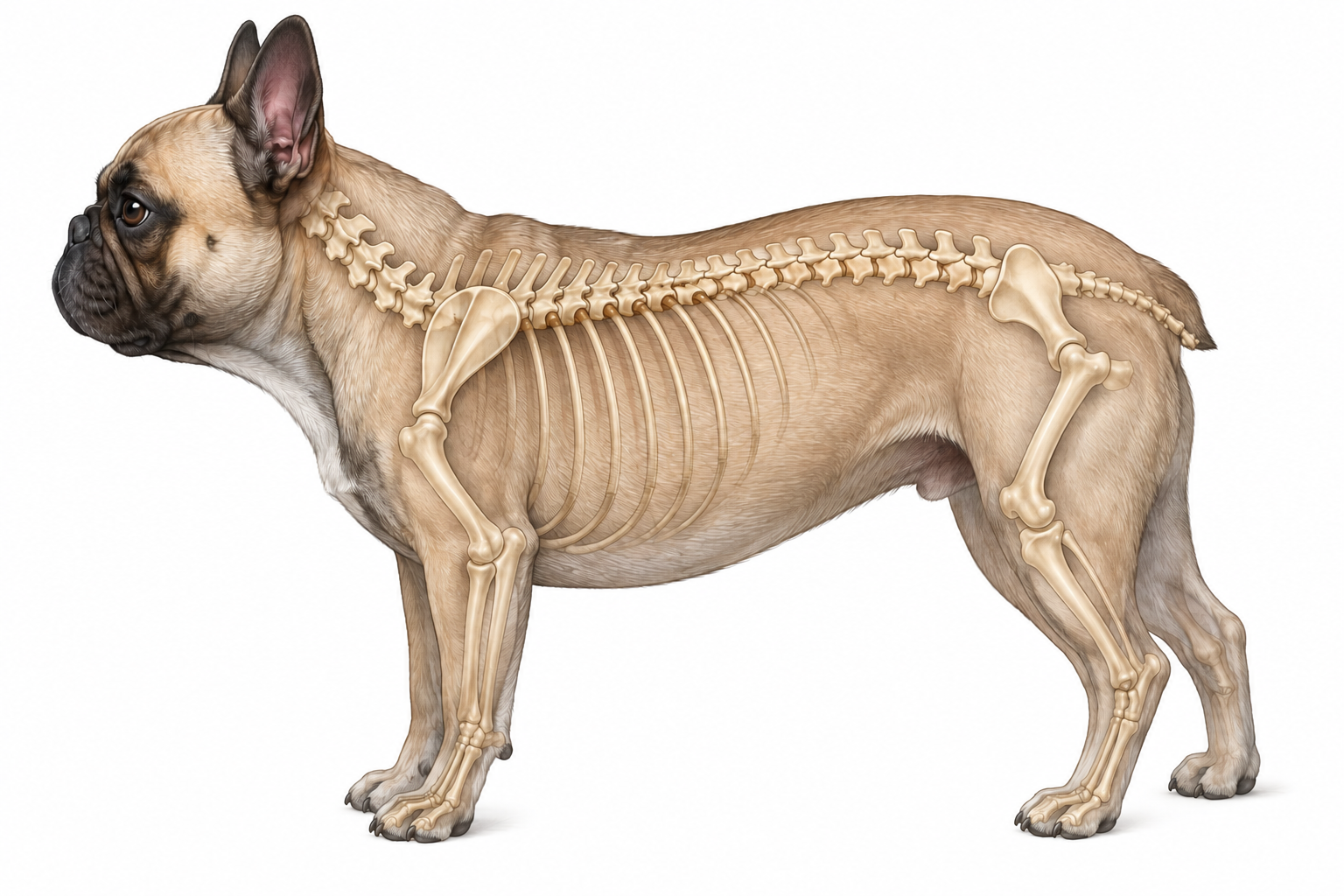
Select a point on the image to view symptom details.
IVDD Grades
The severity of IVDD is generally classified into five neurological grades. This classification helps the vet assess the prognosis and choose the most appropriate treatment.
- Grade I
Pain only
The dog feels pain in the back or neck but continues to walk normally. No obvious neurological deficits are present.
- Grade II
Weakness and ataxia
The dog can still walk but shows an unsteady gait, limb weakness, and difficulty with coordination.
- Grade III
Loss of the ability to walk
The dog can no longer support itself on its legs but retains the ability to move its limbs voluntarily.
- Grade IV
Paralysis with intact deep pain
The dog is paralysed but still perceives deep pain. This is an urgent situation requiring immediate neurological assessment.
- Grade V
Paralysis without deep pain
In addition to paralysis, deep pain perception is absent. This represents the most severe neurological grade and constitutes a true veterinary emergency.
'Deep pain perception' refers to a conscious response to a deep nociceptive stimulus, assessed by the veterinarian during the neurological examination. It should not be tested by the owner at home.
Good to know
Neurological grading is established exclusively by the vet following a complete neurological examination. Two dogs with the same disc herniation may present different clinical grades depending on the degree of spinal cord compression and the time elapsed since the onset of symptoms.
Diagnosis
The diagnosis of IVDD is not based on a single test. The vet collects the dog's clinical history, performs an orthopaedic examination, and above all a thorough neurological examination to identify the location of the lesion and assess its severity.
When spinal cord compression is suspected, imaging studies may be needed to confirm the diagnosis and plan the most appropriate treatment.
- 01
Symptom observation
The vet collects detailed information about when the symptoms appeared, how they have evolved, and any previous episodes. Even small changes in gait or behaviour can help guide the diagnosis.
- 02
Neurological examination
The neurological examination assesses posture, spinal reflexes, limb strength, coordination, pain perception, and the location of the lesion along the spine. This step is fundamental in deciding which further tests are needed.
- 03
Imaging studies
X-rays can reveal abnormalities of the spine but do not directly show spinal cord compression. When necessary, the vet may recommend an MRI or CT scan, which allow the lesion to be located more precisely.
- 04
Neurological severity assessment
Once the lesion has been located, the vet evaluates the severity of the neurological deficit. The ability to walk, limb strength, spinal reflexes, and deep pain perception are all examined. This information is essential for estimating the prognosis and guiding treatment decisions.
- 05
Treatment selection
The information gathered during the neurological examination and diagnostic tests makes it possible to choose the most appropriate treatment. In mild cases conservative management may be indicated, while significant spinal cord compression or major neurological deficits may require surgery.
- 01
Symptom observation
The vet collects detailed information about when the symptoms appeared, how they have evolved, and any previous episodes. Even small changes in gait or behaviour can help guide the diagnosis.
- 02
Neurological examination
The neurological examination assesses posture, spinal reflexes, limb strength, coordination, pain perception, and the location of the lesion along the spine. This step is fundamental in deciding which further tests are needed.
- 03
Imaging studies
X-rays can reveal abnormalities of the spine but do not directly show spinal cord compression. When necessary, the vet may recommend an MRI or CT scan, which allow the lesion to be located more precisely.
- 04
Neurological severity assessment
Once the lesion has been located, the vet evaluates the severity of the neurological deficit. The ability to walk, limb strength, spinal reflexes, and deep pain perception are all examined. This information is essential for estimating the prognosis and guiding treatment decisions.
- 05
Treatment selection
The information gathered during the neurological examination and diagnostic tests makes it possible to choose the most appropriate treatment. In mild cases conservative management may be indicated, while significant spinal cord compression or major neurological deficits may require surgery.
X-ray
Can help identify bone abnormalities, disc calcifications, or other spinal pathologies. On its own, however, it generally cannot confirm spinal cord compression.
CT scan
CT provides detailed images of bony structures and is particularly useful in some clinical situations. It can be combined with myelography or used when MRI is not available.
MRI
MRI is the reference examination for assessing the spinal cord and precisely locating the compression caused by disc herniation. The information obtained helps the vet choose the most appropriate treatment.
Good to know
Not all dogs immediately require advanced imaging. The choice depends on the severity of symptoms, the neurological examination, and the patient's clinical condition.
Treatment
The choice of treatment depends on the severity of the neurological deficit, the speed of progression of symptoms, and the dog's general condition. In some cases conservative management is sufficient, while in others timely surgery may be necessary.
Both approaches require close veterinary supervision and, in surgical cases, a structured rehabilitation programme to support the recovery of neurological function.
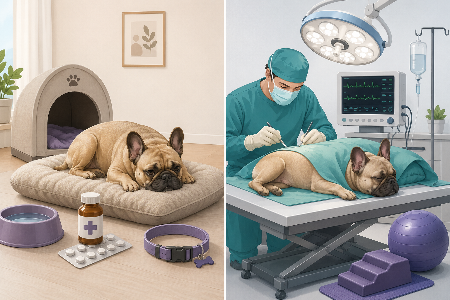
Select a point on the image to view details.
Conservative treatment
- Strict rest in a safe and restricted space — for example a crate or a small controlled area — as directed by the veterinarian. The size, duration, and nature of the restriction must be adapted to each individual case.
- Anti-inflammatory and analgesic medications prescribed by the vet
- Non-slip surfaces and an obstacle-free environment
- No jumping, no stairs, no strenuous physical activity
- Regular veterinary check-ups to monitor progress
- Gradual return to activity only on veterinary advice
Surgical treatment
- Indicated for severe or rapidly worsening neurological deficits
- Most common technique: hemilaminectomy to decompress the spinal cord
- The timeliness of surgery influences the prognosis
- Essential when deep pain perception is absent
- Structured post-operative rehabilitation for recovery
- Early treatment may improve recovery prospects, but outcomes depend on multiple factors assessed by the veterinarian
Good to know
The choice between conservative and surgical treatment does not depend solely on symptom severity, but also on the speed of neurological deterioration and the response to therapy. In some cases the vet may recommend surgery even without complete paralysis, if the clinical picture is worsening.
⚠️ When every minute counts
The sudden loss of deep pain perception in the limbs is a neurological emergency. In this situation, every hour can significantly affect the possibility of recovery. Contact a veterinary centre with neurosurgical availability immediately.
Recovery and prognosis
Recovery after an episode of IVDD varies from dog to dog and depends on numerous factors, including the severity of neurological damage, the speed of diagnosis, the chosen treatment, and the individual response to therapy.
Some dogs recover completely, while others may retain permanent neurological deficits or require a longer rehabilitation period. For this reason, it is essential to carefully follow the veterinarian's instructions throughout the entire recovery process.
Many French Bulldogs, especially when diagnosis and treatment are prompt, can recover a good quality of life. However, every case is different and recovery depends on the severity of neurological damage, the speed of intervention, and the individual response of the dog.
- 01
First hours and days
After diagnosis, the main goal is to manage pain, limit further neurological damage, and closely monitor clinical progression.
- 02
First weeks
Controlled rest and adherence to the veterinarian's recommendations are a fundamental part of recovery. During this phase, periodic clinical check-ups may be scheduled.
- 03
Gradual return to activity
When the veterinarian considers it appropriate, the dog may begin a progressive return to activity and, in indicated cases, a physiotherapy or veterinary rehabilitation programme.
- 04
Ongoing monitoring
Even after recovery, it is important to continue monitoring the dog and watching for the possible onset of new symptoms, as further episodes may occur.
- 01
First hours and days
After diagnosis, the main goal is to manage pain, limit further neurological damage, and closely monitor clinical progression.
- 02
First weeks
Controlled rest and adherence to the veterinarian's recommendations are a fundamental part of recovery. During this phase, periodic clinical check-ups may be scheduled.
- 03
Gradual return to activity
When the veterinarian considers it appropriate, the dog may begin a progressive return to activity and, in indicated cases, a physiotherapy or veterinary rehabilitation programme.
- 04
Ongoing monitoring
Even after recovery, it is important to continue monitoring the dog and watching for the possible onset of new symptoms, as further episodes may occur.
Initial severity
The severity of neurological symptoms at the time of examination is one of the main factors influencing the prognosis.
Deep pain perception
The presence of deep pain perception is one of the most important prognostic factors assessed by the veterinarian. Its absence makes recovery more uncertain, but the prognosis always depends on the overall clinical picture.
Speed of intervention
Timely diagnosis and treatment can improve the chances of recovery in many cases of IVDD.
Individual response
Each dog responds differently to treatment. Age, general condition, location of the lesion, and severity of compression all influence the recovery process.
Possible recurrences
Treating one episode of IVDD does not eliminate the spinal predisposition. Some dogs may develop new episodes over time, making continuous monitoring and adherence to veterinary recommendations important.
Good to know
Recovery times can vary considerably. Some dogs show improvement within days or weeks, while in cases with significant neurological deficits recovery and rehabilitation may continue for several months. There is no single timeline that applies to all cases: progress must be assessed by the veterinary team overseeing the dog.
Prevention and daily management
In the French Bulldog, it is not possible to eliminate the risk of IVDD entirely. Genetic predisposition and early degeneration of the intervertebral discs can be present even in a young, active, and well-managed dog.
Careful management can, however, help keep the dog in good physical condition, reduce some avoidable stresses on the spine, and promote early recognition of pain or neurological deficits.
Daily measures must be adapted to the individual dog. After an episode of IVDD, surgery, or the onset of neurological symptoms, the activity plan must be established by the veterinarian or the veterinary rehabilitation professional overseeing the patient.
Maintain an ideal body condition
Excess weight increases the load the dog must bear during movement and can make it harder to maintain good mobility. Regularly monitor weight and body condition with your veterinarian, adapting diet and activity to the individual dog's needs.
Limit repetitive jumping
Repeatedly getting on and off sofas, beds, or cars can subject the spine to avoidable stresses. Where possible, use a stable ramp or steps suited to the dog and teach it to use them calmly, without forcing it.
Make the environment safer
Very slippery floors can make it harder to maintain stability and movement control. Non-slip rugs, securely fixed runners, and ramps with an adherent surface can facilitate movement and reduce the risk of slipping.
Ramps should be stable, sufficiently wide, and with a slope suited to the dog. Ramps can reduce some strain on the spine, but they do not eliminate the risk of IVDD and do not replace proper veterinary management.
Prefer regular and controlled activity
Appropriate activity helps maintain muscles, mobility, and body condition. Regular, controlled walks are generally preferable to long periods of inactivity alternated with sudden, very intense exercise. After an episode of IVDD, the amount and type of exercise must be determined by the veterinarian. Do not independently start swimming, treadmill, balance exercises, climbing, stairs, or strengthening programmes.
Avoid sudden pulling
During walks, use properly fitted equipment and manage the lead to avoid sudden jerks. A correctly chosen harness may help distribute forces more evenly compared to traction concentrated on the neck, but does not prevent IVDD and must be comfortable and well adjusted.
Recognise the first signs promptly
Stiffness, reluctance to move or jump, a hunched back, pain when the dog is lifted, uncertain steps, nails scraping the ground, and limb weakness must not be ignored. Stop the activity and contact your veterinarian if symptoms compatible with a spinal problem appear.
Review IVDD symptomsWhat not to do
Certain behaviours can delay diagnosis or interfere with recovery.
Do not manipulate the back
Do not attempt deep massage, vertebral "unlocking", traction, or spinal manipulation without a diagnosis and without veterinary recommendation.
Do not administer medication independently
Do not use pain relievers, anti-inflammatories, or other human or veterinary medicines without a specific prescription. Certain combinations can be dangerous.
Do not force movement
If the dog shows pain, weakness, or difficulty walking, do not force it to walk, climb stairs, or perform exercises to see if it "gets better".
Do not wait for an obvious worsening
Neurological deficits can progress rapidly. Intense pain, loss of coordination, dragging of paws, or inability to walk require timely veterinary assessment.
After an episode of IVDD
After conservative or surgical treatment, the return to normal activity must be gradual and guided by the veterinary team. Even when the dog appears to be doing well, the tissues may not have completed recovery.
- 1
Respect the prescribed activity restriction period.
- 2
Perform only the exercises indicated by the veterinarian or the veterinary rehabilitation professional.
- 3
Keep ramps, non-slip surfaces, and other environmental modifications in place even after improvement, if advised.
- 4
Report immediately any recurrence of pain, weakness, instability, or paw dragging.
Good to know
Correct management can reduce some avoidable stresses, but cannot eliminate disc degeneration or guarantee that an episode of IVDD will not occur. The aim is to keep the dog in good condition, adapt the environment to its needs, and act promptly when symptoms appear.
When IVDD is an emergency
IVDD can evolve very differently from one dog to another. Some dogs present only pain, while others can rapidly develop significant neurological deficits.
Recognising the signs that require an urgent visit helps reduce the risk of delays in diagnosis and treatment.
Contact your veterinarian as soon as possible
These symptoms warrant veterinary assessment within the first available hours, even if the dog is still able to walk.
- Neck or back pain
- Stiffness
- Reluctance to jump
- Difficulty climbing stairs
- Unusual posture
- Mild weakness
- Unsteady gait
- Sudden changes in behaviour due to pain
Even if symptoms appear mild, it is advisable to avoid jumping, running, and play until the veterinary visit.
Go immediately to a veterinary clinic
These signs may indicate severe neurological compression and require urgent veterinary assessment.
- Sudden loss of the ability to walk
- Paralysis of one or more limbs
- Marked dragging of the paws
- Inability to stand
- Very rapid worsening of symptoms
- Loss of bladder or bowel control
- Severe pain that prevents the dog from moving normally
What to do while waiting
If you suspect a spinal problem, the aim while waiting for veterinary assessment is to avoid unnecessary movement and not to worsen the situation.
- Limit your dog's movement to the absolute minimum. Do not allow it to run, jump, or use stairs.
- If you need to move the dog, try to keep its body as stable as possible and support both the front and back at the same time. If the dog cannot walk, ask the clinic for guidance on the safest way to transport it.
- Keep the dog on a stable, non-slip surface in a quiet environment.
- Do not give human medicines or any other medications without veterinary guidance. Common human painkillers can be dangerous for dogs.
- Note when the symptoms started and whether the dog is still able to walk, urinate, and move its limbs. This information can be useful for the veterinarian.
Good to know
The severity of IVDD does not depend solely on the intensity of pain. Some dogs can present significant neurological deficits without vocalising or showing obvious pain. For this reason, the ability to walk and neurological status are fundamental elements of the veterinary assessment. During recovery, contact your veterinarian again if the dog shows worsening pain, returns to having difficulty walking, loses limb strength, or new neurological symptoms appear.
Do not wait until the next day
If the dog suddenly loses the ability to walk, develops paralysis, or shows rapid neurological deterioration, contact a veterinarian or an emergency clinic immediately. In many cases, the speed of assessment can influence the chances of recovery.
Can I wait a few days to see if it gets better?
In cases of mild pain without neurological deficits, it is still advisable to have the dog examined as soon as possible. If weakness, loss of ambulation, paralysis, or other significant neurological signs appear, it is not advisable to wait and see how symptoms evolve spontaneously.
Frequently asked questions
The following answers provide general information based on current veterinary knowledge and do not replace a clinical examination.
It depends on the severity of symptoms, the neurological examination findings, and diagnostic test results. Some dogs can be managed with conservative treatment, while others require surgery. The choice is made by the veterinarian on a case-by-case basis.
Many dogs recover a good quality of life; some recover full normal function, while others may retain residual neurological deficits or develop new episodes over time. Recovery depends on the severity of the injury, the initial neurological status, the treatment received, and the dog's individual response.
Return to activity must be gradual and determined by the veterinarian. After recovery many dogs can resume a normal family life, but some adjustments may be needed to reduce strain on the spine.
No. Ramps can reduce some of the stress associated with repeated jumping, but they do not eliminate the genetic predisposition or reliably prevent the disease from developing.
Yes. A dog that has already had an episode of IVDD may develop further episodes during its lifetime. For this reason it is important to continue following the veterinarian's advice and to monitor for any new symptoms.
Severe pain, loss of the ability to walk, paralysis, rapid worsening of symptoms, or loss of bladder and bowel control all require urgent veterinary evaluation.
Rest is an important part of IVDD management, but the duration and approach must be determined by the veterinarian for each individual case. Return to activity should be gradual and follow the advice received.
Keeping the dog in good body condition, limiting avoidable spinal stress, using ramps where appropriate, and recognising symptoms early can help with day-to-day management. However, no measure completely eliminates the risk of IVDD.
Rehabilitation can be very beneficial for some dogs with IVDD, but the type of exercise and when to start depend on the neurological picture and the treatment received. At home, perform only the exercises prescribed and demonstrated by the veterinarian or veterinary rehabilitation professional overseeing the dog. Do not independently start movement programmes, stretching, balance exercises, or strengthening routines.
Scientific sources
The content of this page was prepared by consulting guidelines, veterinary neurology texts, and authoritative academic resources. The information is periodically reviewed to keep it up to date.
Content updates
FrenchieCarePro periodically updates its content based on the best available scientific evidence and leading international veterinary guidelines.
Veterinary disclaimer
The information on this page is for educational purposes only and does not replace veterinary consultation, diagnosis, or treatment. If your dog shows symptoms consistent with IVDD or other neurological conditions, contact your veterinarian promptly.