Cherry Eye in the French Bulldog
Prolapse of the gland of the third eyelid
Cherry eye is the prolapse of the gland of the third eyelid, an important structure for the ocular surface and for the production of the tear film. In the French Bulldog the predisposition is documented and the condition frequently appears in young dogs.
It is not only an aesthetic problem: the gland serves an important function and, when possible, the goal of treatment is to preserve it.
At a glance
What it is
Prolapse of the gland of the third eyelid, which normally remains hidden from view.
Who is most commonly affected
Particularly associated with young dogs and certain predisposed breeds, including the French Bulldog.
Treatment principle
When surgical intervention is necessary, the modern objective is generally to reposition and preserve the gland, not to remove it.
Cherry Eye: much more than a "red cherry" in the eye
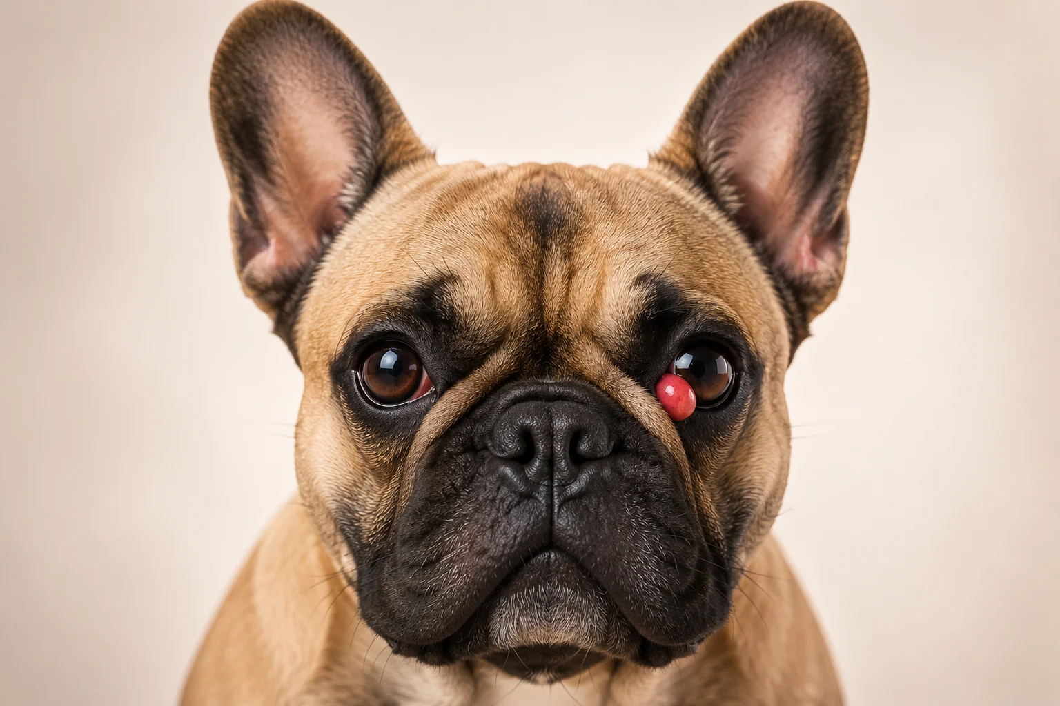
The name Cherry Eye describes the characteristic appearance of the prolapse of the gland of the third eyelid: a smooth, pink or reddish mass that becomes visible in the inner corner of the eye.
The third eyelid, also called the nictitating membrane, is a normal structure of the dog's eye. Associated with it is a lacrimal gland that contributes to the production of the aqueous component of the tear film and therefore to the health of the ocular surface.
Normally the gland remains hidden from view. When the structures that help keep it in position can no longer retain it adequately, the gland may protrude and become visible: this is what is commonly called Cherry Eye.
The French Bulldog is among the breeds in which this condition is observed more frequently. A large VetCompass study conducted on the canine population of the United Kingdom found an annual prevalence of 1.22% in the French Bulldog and a significantly greater predisposition compared to crossbred dogs. The same study also showed a strong association between cherry eye, young age, and brachycephalic conformation.
The fact that cherry eye is easily visible does not mean that every red mass in the corner of the eye can be identified with certainty by the owner. Veterinary assessment is important both to confirm the diagnosis and to check the overall condition of the eye and ocular surface.
Why do we discuss it in the French Bulldog?
annual prevalence observed in French Bulldogs in the VetCompass study
In the large VetCompass study published in 2022, cherry eye was identified in 200 French Bulldogs out of 16,397 included in the analysed population, with an annual prevalence of 1.22%.
In the analysis of the same study, the French Bulldog showed significantly greater odds compared to crossbred dogs. Brachycephaly, considered independently of the individual breed, was also strongly associated with the condition.
These data do not mean that every French Bulldog will develop cherry eye, but confirm that the breed deserves particular attention when a compatible mass appears in the inner corner of the eye.
What exactly is the third eyelid?
To understand cherry eye, we first need to look at a structure of the eye that normally goes almost unnoticed: the third eyelid.
In addition to the upper and lower eyelids, dogs possess a third eyelid, also called the nictitating membrane. It is located in the inner corner of the eye, close to the nose, and is lined with conjunctiva.
It is not a useless or accessory structure. It participates in protecting the ocular surface, helps to spread the tear film, and contains a gland that is important for the production of the aqueous component of tears.

A structure that normally stays mostly hidden
Under normal conditions, the third eyelid sits in the inner corner of the eye and most of its structure remains barely visible.
It consists of a fold lined with conjunctiva and contains an internal cartilaginous support structure, anatomically described as T-shaped cartilage.
The third eyelid contributes to protecting the eye and distributing the tear film across the ocular surface. Its position can become more visible in various ocular conditions, but a visible third eyelid does not necessarily mean cherry eye.
Visible third eyelid ≠ Cherry Eye
In cherry eye it is the gland associated with the third eyelid that prolapses. The third eyelid itself can become more visible for other reasons. The appearance of the eye should therefore be assessed in the context of a veterinary evaluation.
The gland that normally stays out of sight
Associated with the third eyelid is a lacrimal gland located deep at its base. Under normal conditions this gland remains hidden and should not appear as a mass in the corner of the eye.
Its function is important: it contributes to the production of the aqueous component of the tear film, which keeps the ocular surface adequately lubricated.
Estimates reported in the veterinary literature are not entirely uniform, but indicate that the gland of the third eyelid provides a substantial share of aqueous lacrimal production. This is precisely why modern management of cherry eye aims, where possible, to preserve the gland rather than remove it.
An important part of tear production
What changes when the gland prolapses?
The gland is normally held in place by supporting structures. In cherry eye, this support can no longer keep it correctly in its position, and the gland shifts, becoming visible beyond the margin of the third eyelid.
Externally it typically appears as a smooth, pink or red mass in the inner corner of the eye. Once exposed, the gland can become swollen, congested, and inflamed.
In some dogs the prolapse may initially appear to reduce or disappear for a period, then recur. This does not necessarily mean that the problem has resolved.
Normal position
The gland remains hidden at the base of the third eyelid and contributes to normal tear function.
Cherry Eye
The gland shifts from its normal position and becomes visible as a pink or reddish mass in the inner corner of the eye.
Why preserve the gland?
The gland of the third eyelid makes an important contribution to tear production. Its removal can increase the risk of insufficient tear production and keratoconjunctivitis sicca (KCS, commonly called dry eye).
For this reason, when surgical treatment of cherry eye is necessary, the modern approach generally aims to reposition and preserve the gland.
PREDISPOSITION
Why is the French Bulldog predisposed to cherry eye?
Cherry eye can appear in dogs of different breeds, but it does not occur uniformly across the canine population. Some breeds are affected much more frequently than others, and the French Bulldog clearly falls among those predisposed.
Predisposition does not mean that every French Bulldog will develop the condition. It indicates that, compared to many other dogs, the breed has characteristics that make prolapse of the gland of the third eyelid more likely.

There is no single cause
Cherry eye is not generally regarded as the result of a single external cause. The central mechanism is related to the inability of the supporting tissues to keep the gland of the third eyelid stably in its normal position.
When this support is insufficient or laxer, the gland can shift from its location and protrude beyond the margin of the third eyelid.
The strong predisposition observed in certain breeds suggests that anatomical conformation and hereditary factors may contribute to individual susceptibility. However, the precise mechanism has not been fully defined, and it is not correct to attribute every case to a single cause.
Breed
Risk varies considerably between breeds. The French Bulldog is among those in which prolapse of the gland of the third eyelid is observed most frequently.
Age
Cherry eye is diagnosed most often in young dogs. Age is therefore an important element of the epidemiological profile of the condition.
Conformation
Epidemiological studies show an association with brachycephalic conformation. However, skull shape alone does not explain all cases.
What do the data tell us about the French Bulldog?
The VetCompass study published in 2022 analysed over 900,000 dogs in the United Kingdom to investigate the epidemiology of prolapsed nictitating membrane gland.
In the multivariate analysis, the French Bulldog showed markedly greater odds of diagnosis compared with crossbred dogs. Brachycephalic conformation was also associated with higher odds compared with mesocephalic conformation.
These results support the existence of a genuine breed and conformation predisposition, but must be interpreted as epidemiological associations: they do not allow prediction of whether an individual French Bulldog will develop cherry eye.
odds of cherry eye in the French Bulldog compared with crossbred dogs in the VetCompass study
An odds ratio is not the same as saying that a French Bulldog has a 34.1× absolute probability of developing cherry eye. It is a statistical measure of the association observed in the studied population.
The role of brachycephaly
In the VetCompass study, brachycephalic dogs showed greater odds of cherry eye compared with mesocephalic dogs.
This finding indicates an association between skull conformation and the condition, but does not prove that brachycephaly alone causes the prolapse. Breed, the anatomy of the supporting tissues, and other factors may contribute together to the predisposition.
Why do we often see it in young dogs?
One of the most characteristic features of cherry eye is the young age at diagnosis.
In the VetCompass study, an age of less than one year was strongly associated with the condition compared with dogs aged between two and under four years. More broadly, frequency decreased as age increased.
This helps to explain why many owners encounter the problem when their French Bulldog is still a puppy or very young.
Young age, however, is not an absolute rule: cherry eye can also be diagnosed in an adult dog.
Cherry eye is diagnosed more frequently at this stage of life.
It can also appear in adult dogs, but is less epidemiologically characteristic.
The French Bulldog is not the only predisposed breed
Cherry eye is also described in numerous other breeds. Epidemiological studies show a particularly marked predisposition in some canine populations.
Among the breeds frequently associated with the condition are, among others, the English Bulldog, American Cocker Spaniel, Lhasa Apso, Beagle, and other brachycephalic or specifically predisposed breeds.
The involvement of multiple predisposed breeds reinforces the hypothesis that anatomical and hereditary characteristics play an important role in the development of the problem.
Predisposed does not mean destined to develop it
Owning a French Bulldog does not mean that cherry eye will necessarily appear.
Predisposition describes a greater frequency of the condition in the breed compared with other canine populations. It does not allow accurate prediction of what will happen to an individual dog.
Equally, the development of cherry eye does not automatically imply that the owner made an error in day-to-day management.
SYMPTOMS
How does cherry eye present?
The most characteristic sign of cherry eye is the appearance of a smooth, pink or reddish mass in the inner corner of the eye, near the nose.
What the owner sees is not a new growth that has appeared in the eye: in cherry eye it is the gland of the third eyelid that has moved from its normal position and become visible.
The appearance can be very obvious, but is not identical in all dogs. The gland may be more or less swollen and inflamed, and other associated ocular signs may appear.

Illustrazione educativa.
The typical pink or reddish mass
The prolapsed gland typically appears as a smooth, rounded mass in the inner corner of the eye.
The colour can range from pink to red, and the degree of prominence can vary from dog to dog. When the tissue remains exposed it may become more swollen, congested, and inflamed.
It is precisely this appearance that gave rise to the common name ‘cherry eye’.
The sign to remember
A smooth pink or reddish mass in the inner corner of the eye.
What other signs can appear?
In addition to the visible gland, some dogs may show other ocular changes. Their presence and intensity can vary.
Redness
The conjunctiva and the tissues around the gland may appear more red, especially when the gland is irritated or inflamed.
Tearing
The eye may appear wetter than usual or show increased tearing.
Ocular discharge
In some cases ocular discharge may be present. Its appearance warrants attention as it may accompany irritation or other problems affecting the ocular surface.
Irritation
An exposed gland can become irritated and inflamed. In early cases, however, the dog may show few obvious signs of pain.
Can it affect one eye or both?
Cherry eye can affect one eye or both.
In some dogs the problem is initially unilateral and the other eye may be affected at a different point in time. The presence of the problem in one eye does not therefore mean the other will necessarily develop the same condition.
Unilateral
One eye
Bilateral
Both eyes
Can it go back in and then reappear?
Yes. In some cases the prolapsed gland can temporarily return to a less prominent position and appear to have resolved.
It may, however, protrude again at a later stage. A temporary reduction in the visible mass does not therefore allow the conclusion that the problem has been definitively resolved.
Gland visible
Temporarily less prominent
May reappear
One cherry eye can look different from another
A recently prolapsed gland may appear relatively small and pink, while tissue that has remained exposed and irritated may become more swollen and intensely reddened.
For this reason, images found online or the appearance shown in our illustration should not be used as the sole criterion for establishing a diagnosis.
A red mass in the eye is not automatically cherry eye
Other conditions of the third eyelid and the ocular surface can produce visible changes in the same region.
For example, a deformation of the cartilage of the third eyelid can create a pink or red swelling that may resemble cherry eye.
Veterinary assessment is therefore needed to confirm that the visible structure is indeed the prolapsed gland and to check the overall condition of the eye.
Can cherry eye be present without obvious pain?
Yes. Especially in the early stages, some dogs may show few obvious signs of pain or discomfort even though the gland is clearly visible.
This does not mean that the exposed tissue should be ignored. Over time, irritation, inflammation, and other ocular complications may develop.
If the eye appears painful, veterinary assessment is needed
An eye kept shut or half-closed, obvious pain, persistent rubbing, marked changes to the cornea, or a rapid worsening should not automatically be attributed to cherry eye alone.
These signs may indicate a concurrent ocular problem and require veterinary assessment.
Do not try to push the gland back into the eye
If a mass compatible with cherry eye appears, avoid manipulating it or attempting to reposition it manually.
The ocular surface and the gland are delicate tissues. Veterinary assessment allows the problem to be confirmed and an appropriate management plan to be established.
DIAGNOSIS
How is cherry eye diagnosed?
Cherry eye often has a characteristic appearance and the veterinarian can generally recognise the prolapse of the third eyelid gland through examination of the eye.
The consultation, however, does not only serve to confirm that the visible mass is indeed the prolapsed gland. It is also important to evaluate the third eyelid, the ocular surface, and the overall condition of the eye.
Depending on what emerges from the clinical examination, the veterinarian may also consider certain ophthalmic tests to be indicated.

The first step: observing and evaluating the eye
The veterinarian observes the position and appearance of the third eyelid and verifies whether the visible mass is consistent with prolapse of its gland.
The affected eye and the contralateral eye are also evaluated, with attention paid to the conjunctiva, cornea, and other visible ocular structures.
This assessment is important because a mass in the medial canthus should not automatically be considered cherry eye on the basis of appearance alone.
The pathway may vary depending on the appearance of the eye and the findings of the examination.
The diagnosis is not only about the visible mass
Confirming cherry eye is only one part of the assessment. The veterinarian must also consider the condition of the ocular surface and check whether irritation, inflammation, or other concurrent problems are present.
Schirmer tear test: assessing tear production
The Schirmer tear test is used in veterinary medicine to measure aqueous tear production.
A small strip of specific absorbent paper is gently placed in contact with the conjunctiva, generally inside the lower eyelid, and the amount of paper moistened over a standardised interval allows the veterinarian to assess tear production.
In the context of a problem involving the third eyelid gland, knowing the state of tear production can provide useful information about ocular surface function.
Fluorescein: when corneal evaluation is needed
If the eye examination raises suspicion of a problem with the corneal surface, the veterinarian may use fluorescein.
This diagnostic dye allows defects in the corneal epithelium to be highlighted and is commonly used in the evaluation of corneal ulcers.
It does not serve to directly confirm cherry eye: its role is to evaluate the cornea when there is a clinical indication to do so.
Schirmer Tear Test
Provides a measure of tear production.
Fluorescein
Helps to identify corneal surface defects when the test is indicated.
Not all changes to the third eyelid are cherry eye
A pink or reddish structure visible in the medial canthus may have more than one explanation.
Among the changes that may be confused with gland prolapse are problems involving the cartilage of the third eyelid, which can become deformed or everted and produce a visible prominence.
Veterinary examination allows determination of which structure is involved and whether other ocular changes are present.
Gland prolapse
The gland associated with the third eyelid loses its normal position and becomes visible.
Cartilage abnormality
It is the cartilaginous structure of the third eyelid that shows deformation or eversion.
The two conditions are not synonymous and require correct identification during the consultation.
Why does the veterinarian examine both eyes?
Even when cherry eye is evident in only one eye, the ophthalmic assessment should not be limited to the visible mass.
Comparing both eyes can help the veterinarian to evaluate the third eyelid, the ocular surface, and tear production in the context of the individual patient.
Furthermore, as noted in the previous section, the prolapse may affect one or both eyes.
When may a more in-depth evaluation be needed?
Most cases can initially be assessed by the veterinarian through clinical examination of the eye.
When the appearance is atypical, other ocular changes are present, the problem is recurrent, or the situation requires specialist evaluation, the veterinarian may recommend further assessment or referral to a veterinarian with expertise in ophthalmology.
An accurate diagnosis protects more than the gland alone
The aim of the consultation is not only to confirm cherry eye. Assessing tear production, the cornea, and the ocular surface when indicated allows a more complete picture of eye health to be obtained and the most appropriate treatment to be planned.
TREATMENT
How is cherry eye treated?
The treatment of cherry eye depends on the condition of the gland and the eye, but the main objective is to preserve the function of the third eyelid gland and, when necessary, return it to a more appropriate anatomical position.
This principle matters because the gland contributes to the aqueous component of the tear film and therefore to the health of the ocular surface.
Managing irritation or inflammation may be helpful in some cases, but should not be confused with definitive correction of the prolapse.
The fundamental principle: preserving the gland
The third eyelid gland plays an important role in tear production. For this reason, when surgical treatment is necessary, the modern objective is generally to reposition and preserve it rather than remove it.

Managing irritation and correcting the prolapse are not the same thing
When the gland is exposed it can become irritated, congested, or inflamed. Depending on the condition of the eye, the veterinarian may therefore prescribe medical therapy to manage inflammation or other associated ocular problems.
This management may improve the condition of the tissues, but a reduction in redness or swelling does not necessarily mean that the gland has returned stably to its normal position.
When the prolapse persists or recurs, surgical correction may need to be considered.
Medical management
May be used by the veterinarian to manage inflammation, irritation, or concurrent ocular problems when present.
Correction of the prolapse
When indicated, surgical treatment aims to return and maintain the gland in a more appropriate position while preserving its function.
Reducing inflammation and repositioning the gland are therefore two distinct objectives.
Why is it so important to preserve the gland?
The third eyelid gland contributes significantly to the production of the aqueous component of the tear film.
Tears are essential for keeping the ocular surface lubricated and protected.
For this reason, removal of the gland is not considered the approach of choice when preservation is possible: reducing functional lacrimal tissue may increase the risk of problems associated with insufficient tear production.
Why the aim today is to avoid removing it
In the past, the prolapsed gland could be excised. Today the objective is generally to preserve it, because removing a structure that participates in tear production can compromise the eye's ability to maintain an adequately lubricated surface.
When surgery is indicated, the principle is therefore to correct the position of the gland while seeking to preserve its function.
Several surgical techniques exist
Over time, various techniques have been described for repositioning and stabilising the third eyelid gland.
Some procedures create a pocket in the tissues of the third eyelid into which the gland is repositioned; others use anchoring systems or combined techniques.
However, no procedure should be chosen solely on the basis of an online description. The anatomy of the third eyelid, any cartilage abnormalities, the surgeon's experience, and the characteristics of the individual case all contribute to the choice of technique.
Pocket techniques
Techniques commonly described as ‘pocket’ techniques aim to reposition the gland within a pocket created in the tissues of the third eyelid, preserving the gland.
The term describes a general surgical principle: the practical execution belongs to veterinary surgery.
Anchoring techniques
Other procedures aim to stabilise the gland by anchoring it to appropriate anatomical structures. They may be used in selected situations or as part of combined techniques.
The choice depends on the anatomy and the clinical case.
These designations should not be interpreted as a ranking of techniques. The veterinarian chooses the approach based on the individual patient and their own surgical assessment.
What if a cartilage problem is also present?
In some dogs, gland prolapse may be associated with abnormalities of the third eyelid cartilage.
When present, these abnormalities can influence surgical planning and must be identified during veterinary assessment.
This is one reason why treatment must be tailored to the anatomy of the individual eye.
Can cherry eye recur after treatment?
Yes. No technique can guarantee that the gland will remain permanently in position in every patient.
In some cases recurrence may occur and a further veterinary assessment may be necessary.
The risk depends on several factors, including anatomical characteristics, tissue conditions, the technique used, and the individual case.
A recurrence does not automatically mean that something went wrong
The tissue of the third eyelid and anatomical characteristics vary between patients. Even after correct repositioning, in some dogs the gland may prolapse again.
What if both eyes are affected?
When both glands are involved, the veterinarian assesses each eye and establishes the most appropriate treatment plan.
The fact that cherry eye is bilateral does not alter the fundamental principle: to preserve as much gland function as possible and protect the ocular surface.
What not to do
Do not attempt to push, massage, or manipulate the gland to make it return.
Do not use eye drops, ointments, or other ocular medications without veterinary advice: different products have different indications and contraindications, and a painful eye may also have concurrent problems.
If the eye is painful
An eye kept closed or half-closed, obvious pain, persistent rubbing, corneal changes, or rapid worsening require veterinary assessment, as they should not automatically be attributed to cherry eye alone.
The aim is not to remove the gland, but to preserve its function
Cherry eye is highly visible, but the cosmetic result is not the only goal of treatment.
The third eyelid gland is part of the lacrimal system. When intervention is necessary, preserving this structure and maintaining a healthy ocular surface are central aspects of the modern management of this condition.
AFTER SURGERY
Recovery after cherry eye surgery
After surgical repositioning of the third eyelid gland, an important phase begins: protecting the eye while the tissues heal and closely following the veterinarian’s instructions.
The appearance of the eye in the first days may differ from its appearance before surgery. Redness, swelling, and tearing may be present during the initial healing phase, and their intensity can vary from one patient to another.
For this reason, no single postoperative image can serve as a universal reference for independently judging whether an eye is healing normally.

This image is illustrative
The appearance of the eye after surgery may vary. The illustration primarily shows the importance of protection during recovery and should not be used to assess the degree of postoperative redness or swelling.
How may the eye look during the initial phase?
After surgery, the tissues of the third eyelid and conjunctiva are in the process of healing.
During this period, some degree of redness and swelling may be present. Some dogs may also show increased tearing or ocular discharge.
The presence and intensity of these changes depend on the individual patient, the condition of the eye, and the procedure performed.
What matters is not comparing the eye with a photograph found online, but following the progress indicated by the veterinarian and paying attention to any unexpected changes or worsening.
During healing, the following may be observed
Redness
Some degree of redness of the ocular tissues may be present in the initial phase.
Swelling
The tissues of the third eyelid and conjunctiva may temporarily appear more swollen.
Tearing
The eye may appear wetter or tear more during recovery.
Discharge
Some ocular discharge may be present; the amount, appearance, and evolution should be assessed in the context of the instructions received from the veterinarian.
Protecting the eye from rubbing
After eye surgery it is particularly important to prevent the dog from rubbing the eye with its paws or against furniture, carpets, or other surfaces.
The veterinarian may therefore prescribe the use of a protective collar, often an Elizabethan collar, during the healing period.
Even a brief period of rubbing can traumatise a recently operated eye. The protective device must therefore be used in accordance with the instructions received from the veterinary team.
The medication prescribed by the veterinarian
After surgery the veterinarian may prescribe ocular medication and, depending on the case, other medicines to support the postoperative period.
The drug, frequency, duration, and method of administration depend on the individual patient and the procedure performed.
It is important to follow the prescription received exactly and not to modify or discontinue treatment on one’s own initiative.
Do not automatically reuse old eye drops
A product previously prescribed for another ocular problem may not be appropriate in the current situation. Only use products specifically indicated by the veterinarian for that case in the operated eye.
Administering medication without traumatising the eye
When ocular therapy is prescribed, follow the technique shown by the veterinarian or clinic staff.
Avoid letting the tip of the bottle touch the surface of the eye directly, and do not force the eyelids open if the dog shows pain or strong resistance.
If administering the product proves difficult, ask the veterinary team to demonstrate the most suitable technique again.
Rest and activity
During the postoperative period the veterinarian may recommend some temporary limitations on activity, particularly to reduce the risk of knocks, rubbing, or accidental trauma to the eye.
The duration and level of restriction depend on the individual case.
Follow the instructions received regarding walks, play, and the gradual return to normal activities.
Why the veterinary check-up is important
The postoperative check-up allows the veterinarian to assess the position of the gland, the healing of the tissues, and the condition of the ocular surface.
It is also an opportunity to evaluate any redness, swelling, discharge, or other changes and to decide whether the postoperative management should be modified.
Keep the check-up appointments indicated by the clinic even if the eye appears to be doing well.
Timescales and check-ups depend on the individual case and the veterinarian’s instructions.
When to contact the veterinarian
During the postoperative period it is important to observe the eye and the dog’s behaviour. An unexpected change or worsening deserves attention.
- Obvious or increasing pain
- Eye persistently closed or strongly half-closed
- Persistent rubbing
- Marked or rapidly increasing swelling
- Clearly worsening redness
- Abundant discharge or discharge that changes in a concerning way
- Obvious change in the transparency or appearance of the cornea
- Significant bleeding
- Sudden reappearance of a mass consistent with gland prolapse
- Any change that the veterinarian specifically indicated as a reason for a check-up
If you are unsure whether what you are observing falls within the expected course of recovery, contact the clinic that performed the surgery.
May appear during healing
- ·Some degree of redness
- ·Tissue swelling
- ·Tearing
- ·Possible discharge
Requires attention if marked or worsening
- ·Obvious pain
- ·Significant or increasing swelling
- ·Clearly worsening redness
- ·Obvious corneal changes
- ·Persistent rubbing
- ·Reappearance of the mass
This comparison is intended as a guide and does not replace the specific instructions provided by the veterinarian who operated on the dog.
What if the mass reappears?
As explained in the treatment section, prolapse can recur even after surgical repositioning.
If a mass consistent with the prolapsed gland reappears at the inner corner of the eye, contact the veterinarian for a new assessment.
A recurrence must not be manipulated or pushed manually into the eye.
What to avoid during recovery
Manipulating the gland
Do not try to push or massage a mass that reappears.
Letting the dog rub the eye
Protect the eye in accordance with the veterinary instructions.
Modifying treatment independently
Do not discontinue, substitute, or add ocular products without veterinary advice.
Missing check-ups
An eye that appears externally better can still benefit from the scheduled reassessment.
Recovery is not just about the appearance of the eye
After repositioning of the gland, the aim is to allow the tissues to heal, protect the ocular surface, and keep the gland in the position achieved by surgery.
The protective collar, prescribed medication, home observation, and veterinary check-ups are therefore all part of the same recovery path.
PROGNOSIS
What is the prognosis after treatment?
When the third eyelid gland is successfully repositioned and the ocular surface remains healthy, the prognosis is generally favourable.
However, treatment aims to achieve more than a simple cosmetic improvement. The goal is to keep the gland in an appropriate position, preserve its tear-producing function as much as possible, and protect the health of the eye over time.
As with any surgical procedure, the outcome can vary between patients and recurrence or other complications are possible.
A good outcome means more than the mass no longer being visible
Surgical success also includes maintaining gland function and a healthy ocular surface. The external appearance of the eye matters, but it is not the only parameter to consider.
Gland in position
Tear function preserved
Healthy ocular surface
Favourable functional outcome
Can Cherry Eye recur after surgery?
Yes. Even after correct surgical repositioning, the gland may prolapse again.
The likelihood of recurrence is not the same in all patients and may be influenced by various factors, including anatomy of the third eyelid, tissue condition, degree of inflammation, any cartilage changes, and the surgical technique used.
Recurrence does not automatically mean the surgery was performed incorrectly. It means the gland needs to be re-evaluated to determine the most appropriate management.
What do studies show?
Published studies report different success and recurrence rates depending on the population studied, the technique used, and follow-up duration.
For example, a retrospective study of 353 dogs and 420 eyes reported successful repositioning in 95% of cases and an overall recurrence rate of 5%.
These figures should NOT be interpreted as a universal prediction for every French Bulldog or every surgical technique.
95%
successful repositioning in the study of 353 dogs
5%
overall recurrence in the same study
Retrospective study; results may vary between techniques and patients.
Factors that may influence the outcome
Individual anatomy
The characteristics of the third eyelid and its supporting tissues vary between patients.
Inflammation
A gland that is severely inflamed or has been prolapsed for a long time may present with more altered tissue.
Cartilage
Any deformities of the third eyelid cartilage may affect surgical planning and outcome.
Technique and clinical case
The choice of technique is tailored to the individual patient and may influence the outcome.
What complications may occur?
Complications are not inevitable, but should be considered when assessing the post-treatment course.
Recurrence of prolapse
The gland may become visible again and require re-evaluation.
Persistent irritation or inflammation
The ocular tissues may remain irritated or inflamed and require veterinary attention.
Ocular surface problems
Alterations of the cornea or ocular surface may occur and need to be identified and treated.
Changes in tear production
Tear function may be reduced in some patients and may require monitoring over time.
Why do we keep discussing tear production?
Even after gland repositioning, tear production remains an important aspect of ocular health.
Chronic prolapse, inflammation, and compromise of glandular tissue can affect tear function. Some dogs may also have an independent predisposition to keratoconjunctivitis sicca.
For this reason, when indicated, the veterinarian may monitor tear production during follow-up.
The ocular surface deserves attention over time
Veterinary ophthalmology sources note that dogs with a history of third eyelid gland prolapse may benefit from monitoring of tear production over time, particularly when other predisposing factors are present.
Aesthetic outcome
The prolapsed mass is no longer visible and the eye appears more normal.
Functional outcome
The gland remains preserved, tear production is adequate, and the ocular surface remains healthy.
The second aspect is clinically more important than cosmetic appearance alone.
What to do if Cherry Eye recurs?
If a mass consistent with gland prolapse reappears in the inner corner of the eye, veterinary re-evaluation is recommended.
The veterinarian can assess the position of the gland, the state of the tissues, the ocular surface, and decide whether further treatment is needed.
Do not attempt to manually manipulate or reposition the gland.
Recurrence may, in some cases, require a new surgical procedure. The decision depends on the condition of the eye and the assessment of the veterinarian or surgeon.
What about the other eye?
In predisposed dogs, Cherry Eye may also develop in the contralateral eye, even at a different time.
The fact that one eye has been successfully treated does not change the anatomical predisposition of the other.
This does not mean the second eye will necessarily develop the problem, but any new pink or reddish mass should be assessed in the same way.
In summary: what is the prognosis?
With correct diagnosis, appropriate treatment, and adequate follow-up, many dogs achieve a good outcome after gland repositioning.
However, prognosis must be considered on an individual basis, taking into account ocular surface health, tear production, any changes to the third eyelid, and the possibility of recurrence.
Preserve today to protect the eye over time
Treatment of Cherry Eye is not just about the disappearance of the visible mass. Preserving the gland, monitoring ocular surface health, and recognising any recurrence are all part of the same goal: keeping the eye healthy and functional over time.
PUPPY
Cherry Eye in French Bulldog puppies
Cherry Eye is frequently observed in young dogs, and the French Bulldog is among the breeds with a marked predisposition to prolapse of the third eyelid gland.
Seeing a pink or reddish mass appear in the inner corner of a young Frenchie’s eye is therefore a particularly relevant situation for this breed.
Young age alone, however, does not confirm the diagnosis: any new change to the eye must be assessed by a veterinarian.
Why do we discuss this mainly in young dogs?
Epidemiological data show a strong association between cherry eye and young age.
In the VetCompass study already cited in the predisposition section, dogs under one year of age showed significantly greater odds of diagnosis compared with dogs aged between two and under four years.
This finding indicates an epidemiological association with young age: it does not mean that cherry eye can only occur in puppies, nor that every young French Bulldog will develop it.
Young age = predisposition, not certainty
The fact that cherry eye is common in young dogs does not allow prediction of which puppy will develop the prolapse.
At what age can it appear?
Cherry eye can appear early and is diagnosed with particular frequency in young dogs.
There is no precise age at which it must appear. It may be observed during growth, but young age is an associated factor, not a diagnostic rule.
It is therefore incorrect to expect all cases to begin at the same point in development.
Should you wait for the puppy to grow up?
There is no rule stating that cherry eye should simply be left prolapsed until adulthood is reached.
An exposed gland may become irritated and inflamed, while the timing and type of treatment depends on the condition of the eye and veterinary assessment.
The puppy’s age is therefore one element to consider, but should not be used alone to decide whether to wait or to intervene.
Do not decide based on age alone
The appropriate time for treatment is established by considering the puppy as a whole, the eye, and the characteristics of the prolapse.
Can a puppy undergo gland repositioning?
Young age alone does not determine whether a dog can or cannot undergo a surgical procedure.
When repositioning is indicated, the veterinarian assesses the individual patient, the condition of the eye, the dog’s general health, and relevant anaesthetic considerations before planning the procedure.
In the French Bulldog, anaesthetic assessment deserves particular attention also due to the breed’s brachycephalic characteristics.
Can it start in just one eye?
Yes. The prolapse may initially be present in only one eye and, in predisposed dogs, may also appear in the contralateral eye at a different time.
This does not mean the second eye will necessarily be affected.
If a new mass appears in the other eye, this too should be assessed by a veterinarian rather than being automatically considered identical to the first episode.
One eye affected
Does not imply the other will necessarily be affected too
Predisposition
Monitor both eyes over time
Why is preserving the gland particularly important?
The third eyelid gland contributes to the production of the aqueous component of the tear film.
In the treatment of cherry eye, the modern goal is therefore to preserve this functional tissue as much as possible, regardless of whether the patient is young or adult.
In a puppy, this principle takes on a long-term perspective: the aim is not only to correct the visible mass, but to help maintain a healthy ocular surface throughout the dog’s life.
To learn more about why gland preservation and repositioning is today the preferred approach, see the section on cherry eye treatment.
What if the mass appears and then seems to go back in?
In some cases the gland may appear more or less prominent at different times.
The fact that the mass is not always visible does not, however, allow the conclusion that the problem has resolved on its own.
If you have observed a mass consistent with cherry eye, particularly if it recurs, it is worth showing it to a veterinarian. A photograph taken when the mass is visible can help document what you observed, but does not replace examination of the eye.
I noticed a red lump in my puppy’s eye: what should I do?
Observe without manipulating
Do not push, massage, or rub the mass.
Prevent rubbing
Stop the puppy from repeatedly traumatising the eye while arranging a veterinary assessment.
Document what you saw
If the mass changes or temporarily disappears, a photograph can help show the veterinarian what it looked like.
Have it assessed
The veterinarian can confirm which structure is involved and also check the surface of the eye.
A painful eye deserves closer attention
Cherry eye can be very visible, but marked pain, an eye kept closed or significantly narrowed, persistent rubbing, or visible changes to the cornea should not be automatically attributed to the gland prolapse alone. In the presence of these signs, it is important to contact a veterinarian.
Predisposition does not mean…
…it will definitely happen
Not all predisposed puppies develop cherry eye.
…both eyes will be affected
The second eye may remain unaffected.
…you must wait until adulthood
The timing of management is decided based on the individual case.
In a puppy, treating well means thinking about the eye’s future too
Cherry eye can appear early in the French Bulldog, but young age and predisposition should lead neither to automatic alarm nor to ignoring the problem while waiting for the dog to grow up.
A thorough veterinary assessment makes it possible to confirm the diagnosis, monitor the health of the ocular surface, and choose a management approach that also takes into account gland function in the long term.
PREVENTION
Can Cherry Eye be prevented?
There is no proven method that can guarantee prevention of Cherry Eye in a predisposed French Bulldog.
Prolapse of the third eyelid gland is associated with anatomical and individual breed predisposition factors that owners cannot modify through diet, supplements, eye cleaning, or other routine daily practices.
This does not mean there is nothing to do for eye health: it means distinguishing true prolapse prevention from good ocular management and early recognition of a problem.
Predisposition does not mean owner error
A French Bulldog can develop Cherry Eye even when properly cared for. The appearance of a prolapse does not prove the eye was poorly cleaned, the dog ate something wrong, or the owner neglected a specific preventive measure.
What you can control — and what you cannot
NOT CONTROLLABLE
- ·breed predisposition
- ·individual anatomy
- ·characteristics of supporting tissues
MANAGEABLE
- ·observe the eyes
- ·avoid manipulation
- ·protect the eye from trauma and rubbing
- ·seek veterinary assessment when a problem appears
These measures support good ocular health management, but do not eliminate predisposition to Cherry Eye.
Does cleaning the eyes prevent Cherry Eye?
No. Proper hygiene can be useful for routine care of the periocular area, but there is no evidence that eye cleaning prevents prolapse of the third eyelid gland.
Cherry Eye should therefore not be interpreted as a consequence of poor hygiene.
Can diet or supplements prevent it?
There are no foods or supplements proven to specifically prevent Cherry Eye.
A complete and balanced diet is important for the dog's overall health, but should not be presented as a preventive treatment for third eyelid gland prolapse.
Can massaging the eye prevent it?
Regularly manipulating the third eyelid or the inner corner of the eye in an attempt to prevent Cherry Eye is not advisable.
If a mass compatible with gland prolapse appears, do not attempt to push it back or massage it without veterinary guidance.
Home manipulation does not correct the anatomical predisposition and may further irritate the ocular tissues.
What is NOT proven prevention
Frequent cleaning
Ocular area hygiene does not modify the anatomical predisposition to prolapse.
Supplements
There are no supplements proven to specifically prevent Cherry Eye.
Preventive massage
Manipulating the third eyelid does not eliminate predisposition and should not become a preventive routine.
What you can actually do
Even though predisposition cannot be eliminated, certain habits help protect ocular health and quickly recognise any changes.
Observe the eyes
Learn to recognise the normal appearance of your Frenchie's eyes, so you can more easily notice a new mass, redness, or other changes.
Avoid manipulation
Do not press or massage the third eyelid and do not attempt to reposition a prolapsed gland yourself.
Protect from rubbing and trauma
If the eye is irritated or the dog repeatedly tries to rub it, prevent further trauma and seek veterinary guidance.
Have changes assessed
A new mass at the inner corner of the eye warrants a veterinary assessment to confirm which structure is involved and to evaluate the ocular surface.
Recognising it early does not mean it was prevented
Noticing the prolapse quickly allows the eye to be assessed sooner and the most appropriate management to be discussed with the vet. It does not prevent Cherry Eye from having occurred, but can help avoid an evident problem being ignored or improperly manipulated.
Can it be prevented in the other eye?
If Cherry Eye initially affects only one eye, there is no proven home measure that guarantees preventing its appearance in the contralateral eye.
In predisposed dogs the second eye may be involved at a different time, but this is not inevitable.
The most useful strategy is to observe both eyes and have any new change assessed.
Since Cherry Eye is frequently observed in young dogs, attention to the eyes is particularly useful during the French Bulldog's growth. See: Cherry Eye in French Bulldog puppies
What about breeding selection?
The strong predisposition observed in certain breeds suggests an important component linked to conformation and heritable factors, although Cherry Eye cannot simply be attributed to a single identified gene.
Ocular health and the clinical history of breeding dogs are therefore elements that a responsible breeder may consider in the broader context of health-based selection. See also the section on predisposition .
This does not, however, guarantee that a puppy will not develop Cherry Eye.
If you are choosing a puppy
It is not possible to determine from the appearance of a healthy puppy whether it will develop Cherry Eye in the future.
When choosing a French Bulldog, it is more useful to consider the overall health of the parents and the puppy, and to choose a breeder who is transparent about the health history of their dogs.
No commercial promise can guarantee the future absence of Cherry Eye in a predisposed dog.
If Cherry Eye has already appeared
When prolapse is already present, the goal is no longer to prevent its appearance, but to protect the eye, avoid manipulation, and obtain an appropriate diagnosis and management.
After any surgical treatment, following post-operative instructions helps protect the eye during healing, even if it cannot guarantee the absence of recurrence. See post-operative care for further details.
PREDISPOSITION
CANNOT BE ELIMINATED BY A HOME ROUTINE
but
OBSERVATION + PROTECTION + VETERINARY ASSESSMENT
BETTER OCULAR HEALTH MANAGEMENT
The key point
In the French Bulldog, no routine can guarantee that Cherry Eye will not appear.
The most useful thing you can do is understand the breed's predisposition, observe the eyes without manipulating them, and seek a veterinary assessment when a change appears.
WHEN TO CONTACT THE VET
When to see the vet?
Cherry Eye is often very obvious visually, but how urgent the situation is also depends on how the eye as a whole looks and behaves.
A pink or reddish mass compatible with prolapse of the third eyelid gland should be assessed by a vet to confirm the diagnosis and check the health of the ocular surface.
If marked pain, a closed eye, visible corneal changes, or rapid worsening occur, it is important to seek a more prompt assessment, as a concurrent ocular problem may also be present.
Cherry Eye is not only what you can see
A pink mass in the inner corner of the eye may be compatible with Cherry Eye, but the vet must also consider pain, cornea, conjunctiva, discharge, and the dog's behaviour.
Have it assessed by the vet
If a new pink or reddish mass appears in the inner corner of the eye, even if the dog seems well, it should be checked.
- pink/reddish mass compatible with Cherry Eye
- gland that appears and then seems to go back in
- problem present in one eye only
- new mass appearing in the other eye
- mild redness or tearing without obvious pain
The fact that the dog does not appear to be in pain alone does not confirm that the situation is simple or that it can be ignored.
Contact the vet more promptly if something worsens
A change from the initial appearance of the Cherry Eye may indicate increasing irritation or a concurrent ocular problem.
- obvious increase in redness
- rapidly increasing swelling
- more abundant ocular discharge or discharge that changes significantly
- frequent rubbing of the eye
- worsening compared to previous hours or days
- reappearance of the mass after surgery
Some signs require a prompt assessment
If the eye appears clearly painful or the cornea changes in appearance, do not automatically attribute everything to Cherry Eye.
- persistently closed eye
- marked squinting
- obvious pain
- persistent rubbing that is hard to stop
- opacity, spot, whitish area, or obvious change to the cornea
- significant bleeding
- ocular trauma
- rapid worsening of the eye's appearance
- sudden change in vision or behaviour consistent with visual difficulty
These signs may be compatible with a concurrent ocular problem and require a prompt veterinary assessment.
Is Cherry Eye always painful?
No. Some dogs, especially in the early stages, may show few obvious signs of pain even though the gland is visible.
An exposed gland can, however, become irritated and inflamed; a clearly painful eye should not automatically be considered a normal consequence of Cherry Eye.
When pain, a closed eye, or rubbing are marked, the vet should also assess the possibility of concurrent ocular problems.
Why does the cornea deserve particular attention?
The cornea is normally transparent.
If it becomes opaque, shows a whitish area, an irregular surface, or an obvious change compared to the other eye — for example in the case of a corneal ulcer — it is important to have the dog assessed promptly.
Cherry Eye involves the third eyelid gland, but a dog may also have a concurrent problem with the corneal surface.
What to do while waiting for the appointment?
Avoid handling the mass and prevent, as far as possible, the dog from repeatedly traumatising the eye with its paws or rubbing against surfaces.
Do not apply eye drops, ointments, or ophthalmic products not prescribed for the current situation.
If the mass changes in appearance or temporarily disappears, a photograph can help document what you observed and show it to the vet.
No obvious pain ≠ problem to ignore
A dog may have a prolapsed gland without showing marked discomfort. This does not mean that exposed tissue should be left without assessment.
What if Cherry Eye goes back in on its own?
In some cases the gland may temporarily become less visible or return from the prolapsed position.
This does not allow the conclusion that the problem has been definitively resolved.
If you have observed a mass compatible with Cherry Eye, especially if it recurs, mention it to the vet.
What if it reappears after surgery?
A mass that is again visible after surgical repositioning may represent a recurrence and requires a new assessment.
Do not try to manipulate or push the gland into the eye.
The vet can check the position of the gland, the ocular surface, and determine whether new treatment is necessary. — recovery and post-operative care.
What if it happens to a puppy?
Cherry Eye is frequently observed in young dogs and should not be ignored simply because the French Bulldog is still a puppy.
Young age alone does not determine either the diagnosis or the timing of treatment.
The vet must assess the eye and the individual patient. Cherry Eye in the French Bulldog puppy
PINK MASS
ALSO OBSERVE:
- pain?
- closed eye?
- rubbing?
- clear cornea?
- worsening?
CONTACT THE VET IF SOMETHING CONCERNS YOU
When in doubt, have the eye assessed
Cherry Eye is often recognisable, but an owner cannot assess all ocular structures at home.
A veterinary visit allows the gland prolapse to be confirmed and, above all, the ocular surface to be checked for health.
FAQ
Frequently asked questions about Cherry Eye in French Bulldogs
The most common questions about prolapse of the third eyelid gland, treatment, and recovery.
Not necessarily. Some dogs, especially in the early stages, may show few obvious signs of pain even though the gland is visible. An exposed gland can, however, become irritated and inflamed. If the eye is clearly painful, remains closed or strongly squinting, the dog rubs it persistently, or the cornea changes appearance, it is important to have it assessed by the vet.
When to see the vetIn some cases the gland may temporarily become less visible or appear to go back in. It may, however, prolapse again. A temporary disappearance of the mass does not allow the conclusion that the problem has been definitively resolved.
There is no single answer for every dog. Management depends on the condition of the gland, the eye, and the individual case. When the prolapse persists or recurs, the vet may recommend surgical repositioning to preserve the gland and its function.
TreatmentThe third eyelid gland contributes to the production of the aqueous component of the tear film. Its removal reduces functional lacrimal tissue and may increase the risk of problems related to insufficient tear production. This is why today, where possible, the goal is to preserve and reposition it.
Yes. A recurrence is possible even after correct surgical repositioning. The outcome depends on anatomical factors, tissue conditions, any cartilage alterations, and the technique used. If the mass reappears, a new veterinary assessment is indicated.
PrognosisYes. In some dogs Cherry Eye initially appears in one eye and subsequently in the other. This does not mean, however, that the second eye will necessarily be affected.
Yes, Cherry Eye is diagnosed with particular frequency in young dogs and the French Bulldog is a predisposed breed. Young age is an associated factor, but it does not mean that every puppy will develop the problem.
Cherry Eye in puppiesThere is no universal rule that one must wait until adulthood. The timing of management depends on the condition of the eye, the puppy as a whole, and the veterinary assessment.
There is no proven method that guarantees prevention of Cherry Eye in a predisposed dog. Eye cleaning, supplements, or other home routines do not eliminate the anatomical predisposition. The most useful thing is to observe the eyes, avoid manipulation, and have any changes assessed.
PreventionIt is not advisable to manipulate or attempt to manually reposition the gland without veterinary guidance. The ocular surface and the third eyelid are delicate tissues, and manipulation may further irritate them.
The vet can generally recognise the prolapse through examination of the eye, but the visit also serves to assess the third eyelid, the cornea, and the ocular surface. Tests such as Schirmer or fluorescein may be used when indicated, but they are not specific tests that alone «prove» Cherry Eye.
DiagnosisWith a correct diagnosis, appropriate treatment, and adequate follow-up, many dogs achieve a good outcome. The prognosis depends, however, on the individual case and must consider not only the appearance of the eye, but also the position and function of the gland, tear production, and the health of the ocular surface.
PrognosisThe cost can vary significantly depending on the country, the practice, the type of procedure, the necessary tests, anaesthesia, and the possible presence of other ocular problems. It is not appropriate to indicate a universal figure. The veterinary practice can provide a quote for the individual case.
Cherry Eye alone should not automatically be considered a cause of blindness. However, a prolapsed gland can become irritated, and a dog may simultaneously have other ocular problems. Marked pain, corneal changes, or changes in vision require a prompt veterinary assessment.
The strong predisposition of certain breeds suggests an important component related to conformation and heritable factors, but Cherry Eye cannot be explained simply by a single identified gene. It is more accurate to speak of anatomical and breed predisposition.
Predisposition
Related symptoms
Other diseases in the same category
SOURCES
Veterinary and scientific sources
This guide was developed on the basis of specialist veterinary sources and peer-reviewed scientific literature. The sources listed below are the main reference for the page's content.
References
O'Neill DG, McKendrick IJ, Keddy AK, Church DB, Brodbelt DC (2021). Breed and conformational predispositions for prolapsed nictitating membrane gland (PNMG) in dogs in the UK: A VetCompass study. PLOS ONE, 16(12):e0260538.
DOI: 10.1371/journal.pone.0260538 — PMID: 34899621
Open sourceWhite C, Brennan ML (2018). An Evidence-Based Rapid Review of Surgical Techniques for Correction of Prolapsed Nictitans Glands in Dogs. Veterinary Sciences, 5(3):75.
DOI: 10.3390/vetsci5030075
Open sourceMultari D, Vigo S, Niebauer GW (2016). Pocket technique or pocket technique combined with modified orbital rim anchorage for the replacement of a prolapsed gland of the third eyelid in dogs: 353 dogs. Veterinary Ophthalmology, 19(3):214–219.
DOI: 10.1111/vop.12286
Open sourceGuionnet A et al. (2025). Surgical Correction of Prolapse of Nictitating Membrane Gland Using a Variant of the Pocket Technique: A Retrospective Study on 101 Dogs and 126 Eyes. Veterinary Ophthalmology (published online 26 May 2025).
DOI: 10.1111/vop.70031
Open sourceSebbag L, Sanchez RF (2023). The pandemic of ocular surface disease in brachycephalic dogs: The brachycephalic ocular syndrome. Veterinary Ophthalmology.
DOI: 10.1111/vop.13054 — PMID: 36585820
Open sourceStanley RG, Kaswan RL (1985). Surgical correction of third eyelid prolapse in dogs. Journal of the American Veterinary Medical Association, 186(1):83.
DOI: 10.2460/javma.1985.186.01.83
Open sourceAmerican College of Veterinary Ophthalmologists (ACVO). Cherry Eye (Prolapsed Gland of the Third Eyelid).
Open sourceMerck Veterinary Manual. Nasolacrimal and Lacrimal Apparatus in Animals.
Open sourceCornell University College of Veterinary Medicine. Cherry eye in dogs.
Open source
Medical information
The content of FrenchieCarePro is for informational purposes only and does not replace veterinary diagnosis, examination, or treatment. Decisions regarding your dog's health should be made together with your vet.