Radiography / X-ray
Primarily shows vertebral shape, number and overall spinal alignment.
It shows the spinal cord poorly and cannot by itself prove that a malformation causes neurological signs.
Congenital vertebral body malformations
A hemivertebra is a vertebra that formed incompletely before birth. In French Bulldogs these changes are common on imaging, but they are not automatically a neurological disease. Clinical importance depends on spinal shape, stability, spinal-cord compression and the dog’s signs.
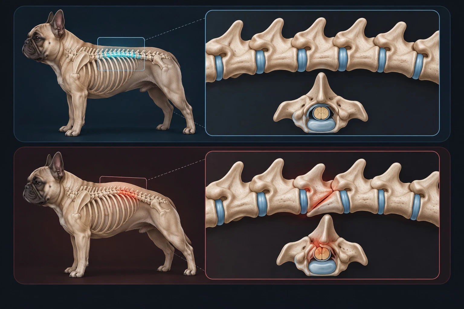
An imaging finding and a clinical diagnosis are not the same thing. A veterinarian must connect the neurological examination with the imaging findings.
A normal vertebra has a broadly symmetrical body, an arch around the spinal canal and joints that guide movement. Incomplete embryonic formation can produce wedge-shaped, butterfly-shaped or other malformed vertebrae. The broader term congenital vertebral malformation is often more accurate because not every abnormal vertebra is a true hemivertebra.
French Bulldogs and other brachycephalic screw-tailed breeds are predisposed. Selection for a short, kinked tail is linked to abnormal vertebral development, but the genetics and clinical consequences are not fully predictable in an individual dog. The thoracic spine, particularly its middle portion, is commonly affected.
A malformed vertebra, spinal deformity, spinal-cord compression and neurological disease are related but distinct findings. A French Bulldog can have several malformed vertebrae and remain neurologically normal.
Malformation can produce kyphosis, scoliosis or rotation. It becomes clinically important when deformity, vertebral subluxation, instability or canal narrowing injures or compresses the spinal cord in a location that matches the neurological examination.
Clinical disease is much less common than incidental imaging findings. When signs occur, they reflect spinal-cord dysfunction rather than bone shape alone and may be progressive, intermittent or occasionally noticed after a minor event.
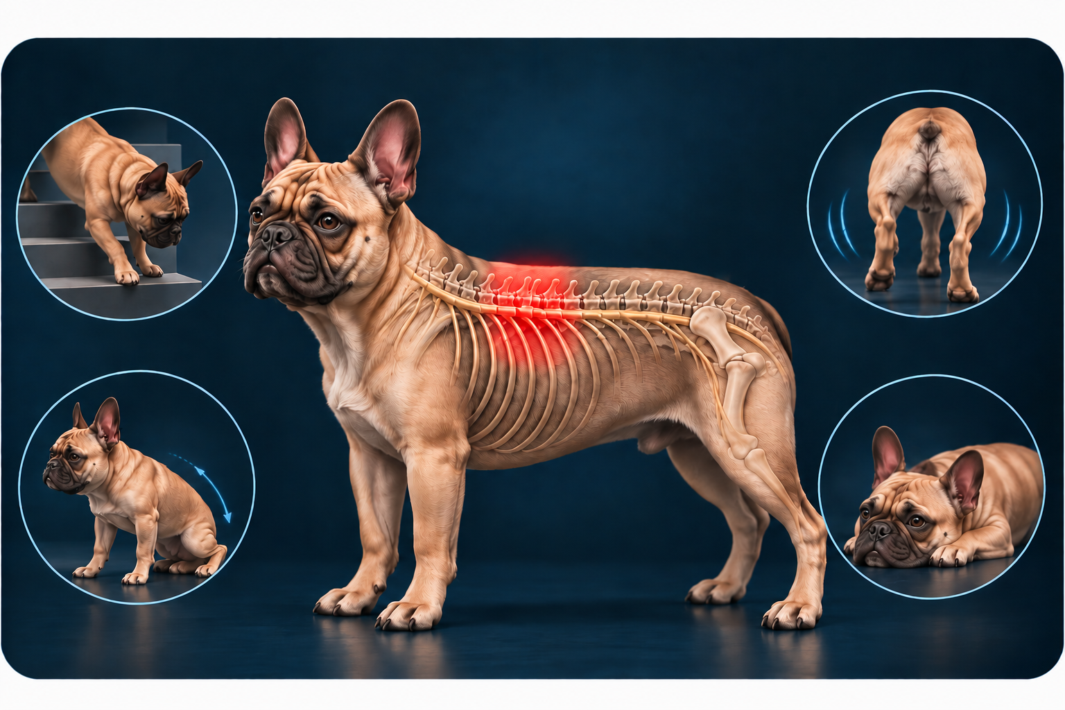
Sudden or worsening weakness, inability to walk, severe pain, loss of bladder control or a rapidly changing gait needs urgent veterinary assessment. Keep the dog as still as practical, support the chest and pelvis during transport and avoid twisting the spine.
These signs are not specific to hemivertebrae. Intervertebral disc extrusion, trauma, infection and other spinal disorders can look similar and may require time-sensitive treatment.
Open the neurological emergency guideAssessment starts with history, general examination and a neurological examination. Gait, postural reactions, spinal reflexes, pain and bladder function help localise a lesion and judge severity. Orthopaedic problems such as patellar luxation must be distinguished from neurological weakness.
Primarily shows vertebral shape, number and overall spinal alignment.
It shows the spinal cord poorly and cannot by itself prove that a malformation causes neurological signs.
Provides detailed cross-sectional views of bone, the vertebral canal and articular anatomy.
It is particularly useful for defining bony anatomy and planning selected surgical procedures.
Best depicts the spinal cord, discs and other soft tissues, including compression or signal change.
Findings still need to match the neurological examination and lesion location.
Radiography, CT and MRI answer different questions and may be complementary. Whatever the modality, imaging becomes clinically meaningful only when the findings match the neurological examination, lesion localisation and the dog’s clinical context.
French Bulldogs are also predisposed to intervertebral disc disease. Other differentials include trauma, discospondylitis, spinal arachnoid diverticulum, meningomyelitis, neoplasia, vascular spinal-cord injury and orthopaedic disease. More than one abnormality can coexist.
A malformed vertebra on an old radiograph should not automatically be blamed for new signs. Lesion location, current examination and advanced imaging must agree.
A neurologically normal dog with an incidental malformation does not automatically need surgery or medication. In one referral imaging study, hemivertebrae were recorded in 93.5% of 62 neurologically normal French Bulldogs. This selected cohort demonstrates how common incidental changes can be; it is not the prevalence in every French Bulldog population.
A veterinarian may recommend documenting the finding, obtaining a baseline neurological examination and monitoring gait, pain and continence. Repeat imaging is not automatic and depends on the individual anatomy, age, breeding plans and any new signs.
Individualised conservative care may be considered for mild, stable signs or when imaging does not support surgery. It may include prescribed analgesia, controlled activity, environmental changes and supervised rehabilitation. No home exercise or supplement can reshape a congenital vertebra.
Strict rest is sometimes used during an acute painful episode, but its duration and the return to activity should be directed by the treating veterinarian. Exercises that force spinal flexion, extension or balance can be harmful when chosen without assessment.
Surgery is reserved for selected dogs with neurological deficits that match a compressive or unstable malformation. The plan may involve stabilisation, realignment and/or decompression and is usually managed by a specialist neurologist or surgeon.
Published evidence consists largely of small retrospective series and case reports. A nine-dog series reported long-term gait improvement in eight dogs after segmental stabilisation, but complications occurred and the sample cannot predict an individual outcome. Risks include neurological deterioration, implant problems, infection and revision surgery.
Prognosis varies widely. An incidental malformation may never cause disease. In clinically affected dogs, outcome depends on severity and duration of spinal-cord dysfunction, continence, cord damage on MRI, stability and whether treatment can address the responsible lesion.
After surgery, recovery usually includes pain control, restricted activity, wound checks, neurological reassessment and staged rehabilitation. Published evidence does not support one universal recovery timeline. New pain, weakness, wound changes or loss of bladder control requires prompt contact with the treating team.
Keep the dog lean and conditioned without high-impact exercise. Excess body fat increases the work of movement and can complicate mobility and anaesthesia, although weight loss cannot correct malformed bone.
Use non-slip flooring, block unsafe stairs when needed and choose controlled regular activity appropriate to the examination. Ramps should be stable and gently sloped. A harness can aid controlled handling; it does not stabilise the spine.
Congenital malformations cannot be prevented after birth. Breeding screening should follow the applicable national breed-club or veterinary scheme and be interpreted by an appropriately qualified veterinarian.
A 2016 study of 105 French Bulldogs estimated substantial heritability for hemivertebra number and grade, supporting selection away from severe malformation. Screening reduces uncertainty but cannot guarantee unaffected or symptom-free offspring. Breeding decisions should avoid ever more extreme screw tails and prioritise function, health and genetic diversity.
Do not diagnose the cause from radiographs alone, manipulate or force the spine, give human painkillers, start strenuous balance exercises or impose prolonged cage rest without veterinary direction. Do not delay because a dog can still wag the tail or take a few steps.
Arrange a veterinary appointment for persistent back pain, altered gait, nail scuffing, repeated stumbling or reduced willingness to move. Seek emergency care for rapid worsening, inability to stand or walk, severe pain, major trauma or new loss of bladder or bowel control.
Percentages apply only to the populations and methods described in each study.
This guide is for educational purposes only and does not replace veterinary examination, diagnosis or treatment. Contact a veterinarian promptly if your dog develops pain, weakness, an altered gait or loss of bladder or bowel control.
Cookie Settings
We use Google Analytics 4 to understand how visitors use this site and improve our content. Analytics cookies are set only if you accept. Declining keeps all features fully functional. Cookie Policy