
Pododermatitis and Interdigital Furunculosis in the French Bulldog
Inflamed paws, interdigital nodules and draining lesions
Pododermatitis means inflammation of the paw; it is a clinical presentation, not one disease. Interdigital furunculosis is a deeper follicular disorder that can produce painful nodules and draining tracts. Similar-looking lesions can have very different causes, so examination and targeted tests matter.
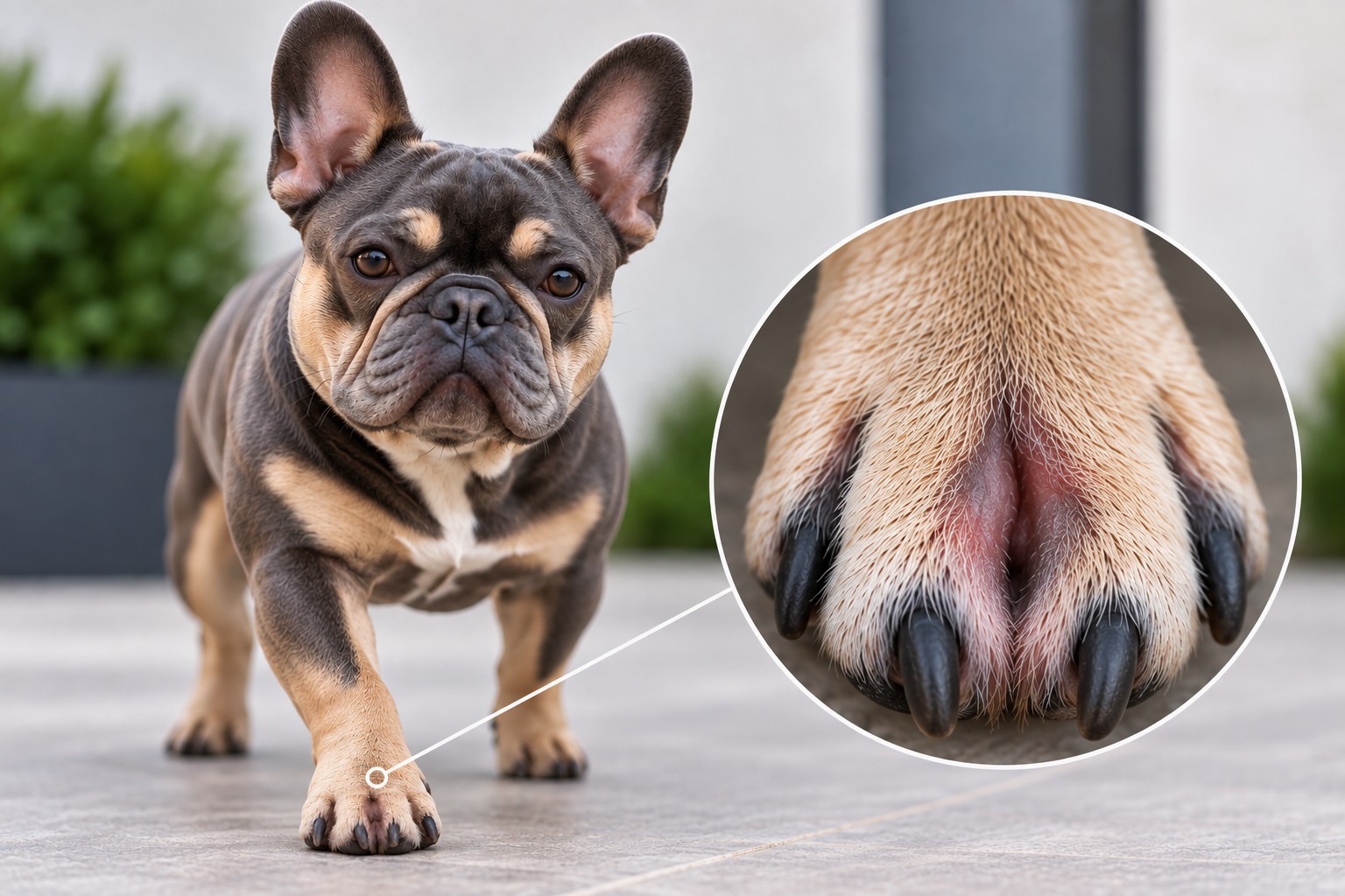
Do not assume every red or swollen paw is ‘an allergy’ or ‘a cyst’. A useful diagnosis identifies the primary cause, any secondary infection, and the factors that keep the lesion recurring.
At a glance
Not one diagnosis
Paw inflammation has allergic, infectious, parasitic, traumatic, immune-mediated and other possible causes.
Depth matters
Surface redness differs from deep nodules, ruptured follicles and draining tracts.
Tests guide care
Cytology, parasite testing and appropriately collected cultures prevent guesswork.
Control the driver
Treating only secondary infection commonly leads to another flare.
Pododermatitis and interdigital furunculosis are not synonyms
Pododermatitis is inflammation of pedal skin and may involve interdigital skin, pads, nail folds or nails. Interdigital furunculosis is deeper inflammation after follicles rupture and release keratin and hair into tissue. The nodules often called ‘interdigital cysts’ are frequently not true epithelial-lined cysts.
A dorsal web may show a nodule or opening, while the weight-bearing palmar or plantar surface may contain comedones, thickening, embedded hairs and continuing follicular damage.
Why it appears: primary, secondary and perpetuating factors
A useful work-up separates what started the inflammation from what now maintains it; more than one factor may coexist.
- Primary causes include allergic skin disease, Demodex or other parasites, foreign material and trauma; less commonly immune-mediated disease, deep fungal or unusual bacterial infection, endocrine disease or neoplasia.
- Predisposing factors can include short coat, body and paw conformation, obesity, abnormal loading, rough surfaces or repeatedly wet feet.
- Secondary bacteria and/or Malassezia yeast can proliferate after barrier damage.
- Licking, ruptured follicles, free hair or keratin, fibrosis, scars, sinus tracts and persistent deep infection can perpetuate disease.
Signs owners may notice
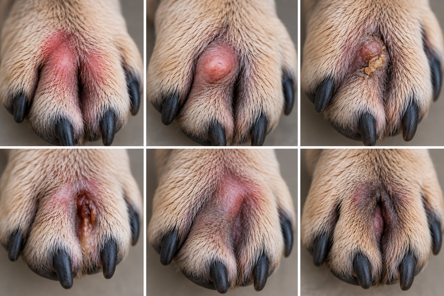
Appearance alone cannot establish cause or depth. Signs range from mild surface inflammation to painful deep disease.
- Repeated licking, chewing or rubbing
- Redness, moist skin, hair loss or brown saliva staining
- Interdigital swelling, papules or firm nodules
- Pustules, crusts, ulcers, blood-tinged or pus-like discharge and draining tracts
- Pain, reluctance to bear weight, lameness or a swollen foot
- Thickened, darkened, scarred skin or recurrence in the same space
One paw versus several: a clue, not a diagnosis
A sudden focal lesion on one paw raises suspicion for trauma, a penetrating foreign body, nail disease, focal deep infection or a mass. Several paws, symmetry, ear disease or itch elsewhere make allergic, parasitic or systemic processes more relevant. These are clues only: allergy may affect one paw prominently, and different secondary infections can occur in different paws.
When veterinary assessment should be prompt
Seek prompt care for marked pain or lameness, rapidly increasing swelling, an embedded object, a deep or draining wound, foul discharge, fever or lethargy, spreading redness, repeated bleeding or deterioration despite treatment. Sudden severe lameness after a walk can fit a grass awn or other penetrating material. Inability to bear weight, major trauma or systemic illness warrants same-day assessment.
See the movement-problems emergency guideDiagnosis: build the answer step by step
History and examination cover onset, seasonality, surfaces, grooming, previous drugs, itch elsewhere, ear disease, gait and all four paws, pads and nails. Tests are selected for lesion depth and distribution.
- Cytology of surface material, exudate or an aspirate to assess inflammation, bacteria and yeast.
- Deep skin scrapings, hair plucks or other parasite tests; fibrotic lesions can make Demodex harder to detect and sometimes require biopsy.
- Careful exploration and selected imaging for suspected foreign material; ultrasound can help in selected lesions but does not replace clinical judgment.
- Culture and susceptibility for relevant deep infection, rods, recurrence, prior antimicrobial exposure or failure; deep tissue is more representative than a superficial swab.
- Biopsy with histopathology, often with tissue cultures, for persistent nodules, draining tracts, unusual lesions or suspected immune-mediated disease or neoplasia.
- Assessment for underlying allergy or systemic disease only when history and full examination support it.
Treatment is guided by cause and depth
There is no universal ‘pododermatitis medicine’. Care may combine pain and itch control, barrier care, parasite treatment, removal of foreign material, allergy management and targeted treatment of confirmed bacterial or yeast overgrowth. Deep furunculosis needs follow-up and may require systemic therapy; chronic scarred tracts or follicular-cyst disease occasionally require referral and surgery.
Current ISCAID guidance supports cytology before antimicrobial use, favours topical treatment for surface and superficial pyoderma, and reserves systemic antimicrobials for appropriate deep or non-responsive cases with reassessment. This guide intentionally gives no drug or dose protocol.
The allergy connection
Atopic dermatitis and cutaneous adverse food reaction can inflame and itch feet, encouraging licking and secondary microbial disease. Dermatological problems are common in French Bulldogs, so allergy is important to consider—but redness, yeast or a nodule does not prove allergy or identify a food. Food allergy requires a properly designed elimination-and-challenge process, not an unvalidated test or rapid food switch.
Why lesions come back
Recurrence usually means a driver remains: uncontrolled itch, altered loading, repeated friction, deep infection, embedded hair or keratin, a missed foreign body, scarring or sinus tracts. A new flare calls for reassessment, not automatic reuse of the previous prescription. Deep disease can persist beneath a quiet-looking surface.
Reasonable home care while awaiting advice
Prevent licking with a well-fitted veterinary collar if tolerated, keep the paw clean and dry, reduce rough-surface activity, and record the affected paw and space. Photograph changes consistently and note pain, discharge and prior treatments. Use a prescribed wash exactly as directed for that dog.
What not to do at home
Do not squeeze, lance or dig into a nodule; probe for a grass awn; apply caustic disinfectants, essential oils or human acne creams; tightly bandage a wet paw; or use leftover antibiotics, antifungals, steroids or painkillers. A poorly fitting, damp boot can worsen friction and maceration.
Outlook and follow-up
Outlook ranges from good for a removable local cause or controllable superficial flare to guarded for chronic, scarred, multifactorial disease. Less redness does not always mean a deep lesion has resolved. Rechecks, repeat cytology or culture when indicated, and long-term control of primary disease reduce avoidable relapse.
Daily risk reduction
Check paws after walks, remove loose surface debris without digging, dry interdigital skin carefully, manage nails and excess hair without injury, and maintain healthy body condition. Choose smooth surfaces during active disease and address persistent licking early. These steps reduce irritation but cannot prevent every case or replace investigation of recurrence.
Frequently asked questions
Scientific sources
This guide synthesises veterinary reviews, consensus guidance and original studies. Evidence specific to French Bulldogs is limited; no breed-specific prevalence is claimed.
- 1. MSD Veterinary Manual (2024). Interdigital Furunculosis in Dogs. MSD Veterinary Manual. Sources
- 2. Bajwa J (2023). Canine pododermatitis: A complex, multifactorial condition. Canadian Veterinary Journal 64:489–492. Sources
- 3. Duclos D (2013). Canine pododermatitis. Veterinary Clinics of North America: Small Animal Practice 43:57–87. Sources
- 4. Duclos DD, Hargis AM, Hanley PW (2008). Pathogenesis of canine interdigital palmar and plantar comedones and follicular cysts, and their response to laser surgery. Veterinary Dermatology 19:134–141. Sources
- 5. Fenet M et al. (2023). Ultrasonographic findings may be useful for differentiating interdigital abscesses secondary to migrating grass awns and interdigital furunculosis in dogs. Veterinary Radiology & Ultrasound 64:920–929. Sources
- 6. Loeffler A et al. (ISCAID) (2025). Antimicrobial use guidelines for canine pyoderma. Veterinary Dermatology 36:234–282. Sources
- 7. Bond R et al. (WAVD) (2020). Biology, diagnosis and treatment of Malassezia dermatitis in dogs and cats: Clinical Consensus Guidelines. Veterinary Dermatology 31:28–74. Sources
- 8. O’Neill DG et al. (2020). Juvenile-onset and adult-onset demodicosis in dogs in the UK: prevalence and breed associations. Journal of Small Animal Practice 61:32–41. Sources
- 9. O’Neill DG et al. (2021). French Bulldogs differ to other dogs in the UK in propensity for many common disorders: a VetCompass study. Canine Medicine and Genetics 8:13. Sources
Veterinary disclaimer
Educational information only. Paw lesions cannot be diagnosed reliably from a photograph and this guide does not replace a veterinary examination. Do not start, stop or reuse prescription, antimicrobial, antifungal, antiparasitic or anti-inflammatory treatment without veterinary direction.