
Skin Fold Dermatitis in French Bulldogs
Intertrigo · facial, nasal and tail-fold inflammation
Friction, trapped moisture and fold anatomy can cause local inflammation. Bacterial or Malassezia overgrowth may become part of the problem, but infection is not present in every case.
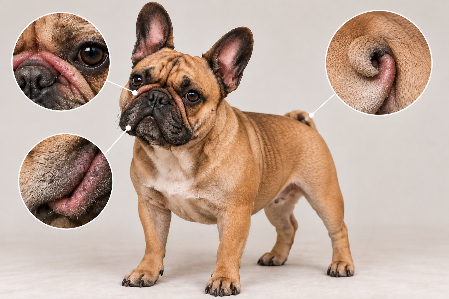
Skin fold dermatitis is a multifactorial inflammatory condition promoted by apposed skin. It is not automatically an allergy, bacterial infection or yeast infection.
In brief
Skin fold dermatitis, or intertrigo, develops where two skin surfaces remain in close contact. Friction, reduced airflow and retained moisture or secretions can disrupt the local skin environment and trigger inflammation. Bacteria or Malassezia yeast may then overgrow, but this does not happen in every case.
French Bulldogs are strongly predisposed because many have prominent facial folds and compact or screw-tail anatomy. In a UK primary-care study, 2.69% had a recorded diagnosis during 2016; this is a one-year recorded prevalence, not a universal or lifetime probability. The adjusted odds were 25.92 times those of crossbred dogs in that study.
Where it occurs
The strongest French Bulldog-specific sites are the facial and nasal folds, including periocular portions, and folds around a short, curved or ingrown tail. A deep tail fold is often called a tail pocket.
Lip, neck or other apposed folds can be affected in individual dogs. Perivulvar fold dermatitis is recognised in dogs generally, but is not a defining French Bulldog manifestation. Eye pain, squinting or ocular discharge must not be assumed to come from the skin alone.
How a fold becomes inflamed
Close skin apposition promotes friction and limits ventilation. Tears, saliva, sebum, exudate or debris may remain trapped; prolonged dampness can cause maceration and barrier disruption. In some dogs, bacterial or Malassezia overgrowth then adds to inflammation and discharge.
The sequence is not rigid and not every case includes microbial disease. Continuing anatomy, moisture and self-trauma can help make disease chronic or recurrent.
Inside the fold: interacting factors
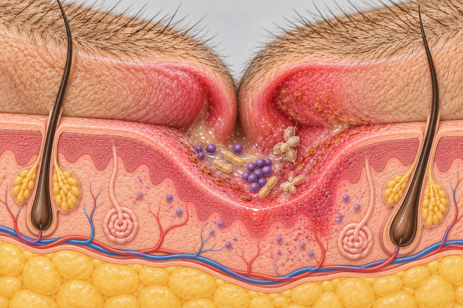
Contact and friction
Opposing skin surfaces remain in contact and rub during normal movement.
Signs to recognise
Early changes may include mild redness, persistent dampness, a new odour, greasy material or sensitivity. More established disease may cause swelling, crusting, discharge, hair loss, rubbing, licking or scooting, pain, erosions or ulceration.
Repeated inflammation can darken and thicken the skin. Odour or redness alone cannot identify an organism or confirm infection.
- Pain, pus-like or bloody discharge
- Open ulcers, draining tracts or rapidly increasing swelling
- Lethargy, reduced appetite or other systemic illness
- Eye pain, squinting or eye redness
Recognising changes in a skin fold
These are independent educational examples, not stages or grades. Appearance alone cannot diagnose skin fold dermatitis or microbial infection.
When to contact a veterinarian
- Arrange an examination when redness, moisture or odour persists or worsens
- the fold is painful
- discharge, bleeding, erosion or swelling develops
- the lesion is close to the eye
- a deep tail pocket cannot be inspected safely
- episodes recur
- or the dog seems unwell.
A mildly damp but otherwise normal-looking accessible fold can be monitored and gently dried. Do not repeatedly manipulate a painful fold or force access into a deep tail pocket.
What else can look similar?
Conditions that can coexist
Atopic dermatitis or adverse food reaction may coexist and worsen itching or microbial overgrowth, but isolated fold disease does not prove allergy. Pododermatitis affects the paws; otitis affects the ear canal. These are distinct conditions even when the same dog has more than one.
Conditions to distinguish
Superficial pyoderma is bacterial infection of the epidermis and follicles; Malassezia dermatitis is yeast-associated disease. Neither is a synonym for intertrigo. Demodicosis, contact irritation, trauma and less common inflammatory disorders may also resemble fold disease.
How veterinarians diagnose it
History and examination establish location, fold depth, pain, distribution and possible contributing disease.
Cytology can show inflammatory cells, cocci, rods and Malassezia and help assess whether microbial involvement is clinically relevant. It does not identify every organism to species, provide susceptibility results or diagnose allergy.
Skin scraping or hair examination may be used when mites are a concern. Biopsy is uncommon for typical intertrigo but may be appropriate for unusual, persistent or ulcerative lesions. Imaging is not routine, although it may help assess severe screw-tail anatomy for surgery or associated neurological signs.
Culture and susceptibility are not required for every active or recurrent lesion. Cytology asks whether microorganisms and inflammation are present; culture identifies cultivable bacteria and their susceptibility. Culture is especially relevant for suspected deep infection, rods on cytology, failure of an appropriate treatment, concern about resistance after relevant antimicrobial exposure, or when systemic antimicrobials are being considered. Recurrence alone does not make culture mandatory.
Treatment and recurrence control
Management addresses inflammation, moisture, microbial involvement and anatomy.
Gentle cleaning and drying may improve the fold environment
topical antiseptic, antimicrobial, antifungal or anti-inflammatory treatment is selected according to examination and cytology.
Recurrent disease may lead the veterinarian to assess allergy, food reaction, mites, wider bacterial or Malassezia disease, dental or ocular problems, body condition and severe tail anatomy. Treating a coexisting disorder may reduce recurrence, but it cannot always remove the mechanical effect of a deep fold.
For suitable surface or superficial bacterial disease, topical antimicrobial management is preferred. Systemic antibiotics are not a routine next step because topical care is inconvenient or improvement is not immediate; they are reserved for selected circumstances after veterinary reassessment of diagnosis and depth. No drug, dose or universal chlorhexidine schedule is provided here.
Home care and prevention
Do
Inspect the folds your dog actually has and learn their normal appearance and smell. In a healthy, accessible fold, gently remove visible moisture or debris with a method approved for that dog and dry without aggressive rubbing. There is no evidence-based schedule for every French Bulldog.
Avoid
Use only products suitable for the skin condition and exact site. Avoid irritating or inappropriate non-veterinary treatments and products whose ocular or mucosal safety is uncertain. Do not place products into an ulcerated fold or a deep, painful or inaccessible tail pocket without veterinary guidance. Appropriate body condition supports general health but cannot remove fixed facial or screw-tail anatomy.
When signs recur
After bathing or getting wet, check folds that tend to retain water and dry them gently. Recurrence is a reason to reassess the diagnosis and plan rather than repeatedly reuse treatment without knowing whether microbes, another skin disease or anatomy are contributing.
When anatomy may require surgery
Most mild or occasional episodes are managed medically. Surgery is considered only for selected dogs when severe facial or screw-tail anatomy repeatedly causes painful disease that cannot be controlled acceptably with medical management.
Options may include removal of diseased fold tissue or partial or complete caudectomy according to anatomy. Evidence comes mainly from small retrospective series and complications can occur. This is therapeutic surgery for significant disease, not routine prevention or cosmetic tail docking.
Many localised cases improve when inflammation, moisture and clinically relevant microbial overgrowth are controlled, but recurrence remains possible because treatment does not necessarily change the fold. The outlook depends on fold depth and accessibility, lesion extent, coexisting disease and whether anatomical correction is needed.
Frequently asked questions
Scientific sources
Curated evidence supporting the guide. Study populations and limitations are stated in the text.
- 1. O’Neill DG, Rowe D, Brodbelt DC, Pegram C, Hendricks A (2022). Ironing out the wrinkles and folds in the epidemiology of skin fold dermatitis in dog breeds in the UK. Scientific Reports 12:10553. Source
- 2. O’Neill DG et al. (2021). French Bulldogs differ to other dogs in the UK in propensity for many common disorders: a VetCompass study. Canine Medicine and Genetics 8:13. Source
- 3. O’Neill DG et al. (2018). Demography and disorders of the French Bulldog population under primary veterinary care in the UK in 2013. Canine Genetics and Epidemiology 5:3. Source
- 4. Paterson S (2017). Intertrigo in the dog: aetiology, clinical signs and therapy. Companion Animal 22(2):72–77. Source
- 5. Banovic F, Strzok E (2019). Skin Fold Dermatitis (Intertrigo) in Dogs. Today’s Veterinary Practice. Source
- 6. Rexo A, Hansen B, Krumbeck JA, et al. (2022). Effect of topical medication on the nasomaxillary skin-fold microbiome in French Bulldogs. Veterinary Dermatology 33(1):10–e5. Source
- 7. Loeffler A, Cain CL, Ferrer L, et al. (2025). Antimicrobial use guidelines for canine pyoderma by ISCAID. Veterinary Dermatology 36(3):234–282 · doi:10.1111/vde.13342 · PMID:40338805. Source
- 8. Knight SM, Radlinsky MG, Cornell KK, Schmiedt CW (2013). Postoperative complications associated with caudectomy in brachycephalic dogs with ingrown tails. JAAHA 49(4):237–242 · doi:10.5326/JAAHA-MS-5858 · PMID:23690492. Source
- 9. Abrescia P, Cinti F, Penazzi C, et al. (2017). Caudectomy for resolution of tail fold intertrigo in brachycephalic dogs: a report of eleven cases. Veterinaria 31(2).
- 10. Mansour TA et al. (2018). Whole genome variant association identifies a DVL2 frameshift mutation in Bulldogs and related screw-tail breeds. PLoS Genetics 14:e1007850. Source
- 11. Miller WH, Griffin CE, Campbell KL (2013). Muller & Kirk’s Small Animal Dermatology, 7th edition. Elsevier, pp. 678–680.
- 12. Gross TL, Ihrke PJ, Walder EJ, Affolter VK (2005). Skin Diseases of the Dog and Cat: Clinical and Histopathologic Diagnosis, 2nd edition. Blackwell, pp. 261–263.
- 13. Lightner BA, McLoughlin MA, Chew DJ, et al. (2001). Episioplasty for the treatment of perivulvar dermatitis or recurrent urinary tract infections in dogs with excessive perivulvar skin folds: 31 cases (1983–2000). Journal of the American Veterinary Medical Association 219:1577–1581.
- 14. Hobi S, Barrs VR, Bęczkowski PM (2023). Dermatological problems of brachycephalic dogs. Animals.
Informational disclaimer
This guide provides general educational information and cannot diagnose an individual dog. Treatment depends on location, lesion depth, cytology and the dog’s wider health. Seek veterinary advice for pain, discharge, ulceration, eye involvement, recurrence or worsening disease.